Oliver Kepp, PhD, INSERM staff scientist, Centre de Recherche des Cordoliers, Co-Director, Institut Gustave RoussyJayne A. Hesley, Applications Scientist, Cellular Imaging, Molecular Devices,
BIOGRAPHY

NOV 12, 2014 8:00 AM PST
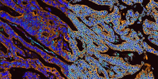
Hallmarks of cancer - Detect and quantify cell death signatures with high content imaging
Abstract
Dr. Oliver Kepp will be presenting on:
Immunogenic cell death fingerprinting utilizing a high-throughput screening approach
The strategy of immunogenic cell death fingerprinting has been designed for the purpose of identifying novel immunogenic anticancer agents in drug re-profiling approaches. Following a general high-throughput screening (HTS) approach (Kepp et al., Nat Rev Drug Discov., 2011) chemical compound libraries have been analyzed by robot-assisted automated microscopy using fluorescent biosensors that were generated for detecting immunogenic cell death signatures (CRT exposure, HMGB1 and ATP release (Zitvogel, Kepp, Kroemer, Cell, 2010)). Using this approach we recently identified compounds from the group of cardiac glycosides, of which some have reached FDA approval for the treatment of heart insufficiency, to be capable of inducing all known hallmarks of immunogenic cell death. The emission of immunogenic cell death factors, triggered by the primary hit compounds, was further validated by low throughput assays and the immunogenic potential of few leads was confirmed in vivo by means of immunocompetent mice and retrospective clinical studies. This successful HTS approach using fluorescent biosensors underlines the capacity of our microscopy-based platform to detect signatures of immunogenic cell death in a large number of different treatment conditions.
Jayne Hesley will be presenting on:
High-content imaging for accurate quantification of autophagy in neuronal cells
Autophagy is a process in which unwanted cellular components, from aged organelles to faulty proteins, are sequestered into double-membrane compartments called autophagosomes. Autophagy is key to maintaining cell homeostasis, as disruption in the process is implicated for conditions such as neurodegeneration, aging, and cancer.
Autophagosomes can be visualized and characterized even in heterogeneous cell populations using readily available reagents, automated imaging and high-content analysis of live or fixed cells. The ImageXpress® Micro XLS system makes it possible to study a large number of treatments with replicates, since it enables acquisition of high resolution, high magnification images at multiple fluorescent wavelengths from cell-based assays run in 384 well microplates. Dose-response curves from human iPSC-derived neurons or PC-12 cells were generated from multiple data outputs, allowing discrimination between simple cytotoxicity and specific effects on cellular function. Accurate co-localization measurements of mitochondria or lysosomes with autophagosomes provide a deeper understanding of the mechanisms of autophagic stimulation or inhibition.
You May Also Like
FEB 08, 2024 | 10:00 AM
High-content screening (HCS) is an imaging-based, multi-parametric strategy used in drug development that generates rich datasets through multiplexing strategically chosen fluorescent dyes a...
OCT 24, 2023 | 8:00 AM

Organoids are three-dimensional (3D) multi-cellular, microtissues derived from stem cells that closely mimic the complex structure and functionality of human organs. They offer more accurate...
OCT 24, 2023 | 10:00 AM

Organoids are three-dimensional (3D) multi-cellular, microtissues derived from stem cells that closely mimic the complex structure and functionality of human organs. They offer more accurate...
NOV 08, 2023 | 8:00 AM
Generating Insights into tissue microenvironments is crucial to our understanding of normal and abnormal tissue development such as during cancer progression. Spatial biology methods using m...
MAR 26, 2024 | 7:00 PM
C.E. CREDITS

The implementation of a preemptive pharmacogenomics (PGx) program in a hospital setting requires a multidisciplinary approach to ensure seamless integration of each stage of the process for...
MAR 26, 2024 | 8:00 AM
C.E. CREDITS
The implementation of a preemptive pharmacogenomics (PGx) program in a hospital setting requires a multidisciplinary approach to ensure seamless integration of each stage of the process for...
Loading Comments...
Finish Registering
-
 APR 30, 2024Immuno-Oncology Virtual Event Series 2024
 -
MAY 07, 20243rd International Biosecurity Virtual Symposium
-
SEP 03, 2024Microbiology Week Virtual Event Series 2024
- See More


-
APR 17, 2024
- See More