Researchers have developed new cancer detection agents in the form of contrast dyes from tattoo ink. The results of these findings are published in the journal Biomaterials Science by a team from USC Viterbi Department of Biomedical Engineering and USC Michelson Center for Convergent Bioscience.
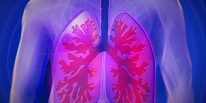
Contrast imaging is a crucial method for detecting cancers. If you’ve ever had an MRI or CT, you may have had a contrast dye injection in order to enhance the precision and sensitivity of the imaging. For cancer, this technique allows doctors not only to diagnose diseases but also provides key images for surgical treatment so that surgeons can remove tumors on the most accurate margins.
"For instance, if the problem is colon cancer, this is detected via endoscopy," explains Cristina Zavaleta, who led the team developing the new contrast agents. "But an endoscope is literally just a flashlight on the end of a stick, so it will only give information about the structure of the colon - you can see a polyp and know you need to take a biopsy. But if we could provide imaging tools to help doctors see whether that particular polyp is cancerous or just benign, maybe they don't even need to take it," she said.
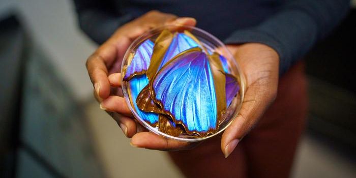
The dyes that Zavaleta’s team has developed come from places so common you might not think to look there: food coloring dyes and tattoo ink. The optical inks, as they are called, are unique because the nanoparticles they’re attached to, which move through the bloodstream to illuminate cancerous tumors, are biodegradable, meaning they’re safe for the human body.

"We thought, let's look at some of the FDA-approved drug, cosmetic and food dyes that exist and see what optical properties are amongst those dyes," Zavaleta said. "And so that's where we ended up finding that many of these FDA-approved dyes have interesting optical properties that we could exploit for imaging."
The key property that the team needed to exploit was the size of the nanoparticles: the dyes need to have nanoparticles small enough to passively penetrate into tumors but not small enough to slip out too quickly before the imaging can be taken.
"With small molecules, you may be able to see them accumulate in tumor areas initially, but you'd have to be quick before they end up leaving the tumor area to be excreted," Zavaleta added. "Our nanoparticles happen to be small enough to seep through, but at the same time big enough to be retained in the tumor, and that's what we call the enhanced permeability and retention effect."
Sources: Biomaterials Science, Eureka Alert