Written By: Eric Torres
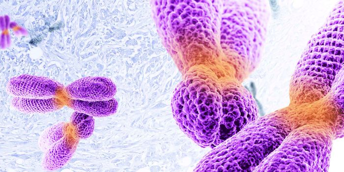
As I pondered the title of this article, my thoughts immediately went back to a request by my Ph.D. advisor to look at the effects on cell viability by an untested drug we suspect inhibits mitochondrial protein import. Cell viability you say? Where to begin…. The myriad of pathways, mechanisms, assays, and associated readouts to measure “viability” quickly made me realize just how comprehensive the term was. For those of you wondering how to get started with cell viability, let’s explore the many tools and metrics to help provide you with some direction.
Are there changes in metabolism or cellular activity?
Assays that measure cellular activity offer the most traditional readouts for viability (often directly called cell viability assays). These assays generally rely on determining relative levels of key metabolic co-factors such as NADH and NADPH by measuring the cell's overall reductive potential. This is done using a molecule, such as MTT or resazurin, that produces a measurable fluorescent or colorimetric output when reduced.
Another metric for cellular activity is the level of ATP, the molecule that provides the energy that drives cellular reactions. The most widely used method for measuring ATP is through a luciferase assay. In this method, cell lysate containing ATP is combined with luciferase and its substrate D-Luciferin to produce a luminescence signal. Biotium’s ATP-Glo™ Bioluminometric Cell Viability Assay is a flash-type luminescence assay that functions this way and can be used for highly sensitive detection of ATP.
Cell viability can also be assessed by monitoring the cell proliferation rate of the culture. CFSE and ViaFluor® stains are membrane-permeant dyes that are non-fluorescent until they enter viable cells where cytoplasmic esterases hydrolyze them to release fluorescent amine-reactive dyes. The dyes then covalently react with amine groups on intracellular proteins and are retained in the cell. Each subsequent cell division reduces the fluorescent signal by half and can be monitored by flow cytometry.
Are my cells undergoing apoptosis?
Assays that are specific for apoptotic activity measure various hallmarks of apoptotic cells and offer a more detailed understanding of cell viability and the mode of cell death.
Measuring the activity of specific apoptotic proteases generally involves end-point assays that rely on a substrate that fluoresces after being cleaved by a specific protease. Biotium’s NucView® Caspase-3 Substrates are a similar type of assay. However, unlike other fluorogenic protease substrates, NucView® Caspase-3 Substrates release a high-affinity DNA binding dye that is retained in the nucleus. This allows real-time monitoring of both caspase-3 activity and changes to nuclear morphology.
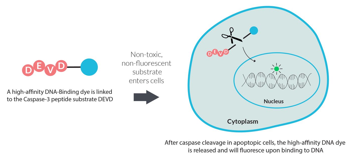
Principle of NucView® substrate technology.
Depolarization or loss of mitochondrial membrane potential is an early event in apoptosis. Consequently, assays monitoring mitochondrial potential are used as a key metric for assessing cell viability. These types of assays rely on cationic dyes that preferentially localize in the matrix of polarized mitochondria. Examples of cationic dyes include MitoView™ 633 as well as classic dyes like TMRM, TMRE, and JC-1.
Another indicator of apoptosis is the externalization of phosphatiylserine (PS) on the surface of the plasma membrane. Annexin V is a cellular protein that binds to PS and is commonly used as a probe for monitoring apoptosis. Fluorescently labeled Annexin V can be used with dead cell dyes like PI or EthD-III for dual detection of apoptosis and necrosis, but can also be used with other cell viability probes.
Internucleosomal dsDNA breaks are another key marker of apoptosis. Apoptosis-induced DNA breaks in fixed cells and tissues can be detected using the TUNEL (terminal deoxynucleotidyl transferase-mediated dUTP nick-end labeling) assay. This assay involves a TdT enzyme that catalyzes the addition of labeled dUTP to the 3’-ends of DNA double-strand breaks. The labeled cells can then be detected with microscopy or flow cytometry.
Are my cells dead? Cytotoxicity or Membrane Integrity Assays
An alternative way to determine the number of live cells in a population is to measure the number of dead cells. Because the loss of membrane integrity is a key indicator for cell death, cytotoxicity assays (aka live/dead assays) rely on membrane-impermeant dyes or substrates to be excluded outside of living cells. Biotium’s Live-or-Dye™ stains as well as all dead cell specific nucleic acid stains rely on membrane integrity to segment live and dead cell populations.

Principle of Live-or-Dye™ discrimination of live and dead cells.
Conclusions
Hopefully, this article provides some insight on the mechanisms behind the various types of cell viability assays to help your research. For more information on cell viability, please see the full article on Biotium’s blog page. You can also visit the Biotium website to learn more about their full selection of cell viability and apoptosis reagents, as well as stains and probes for a variety of cellular structures.