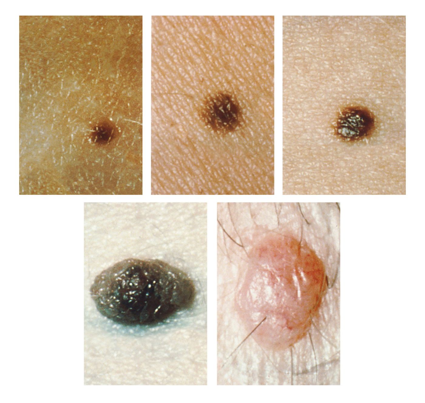
Melanocytes describe a specialized type of cell that generates a substance called melanin, a pigment responsible for skin and eye color. When several melanocytes cluster together, they form a benign, non-cancerous skin growth called a common mole or a melanocytic nevus (plural = nevi). Common moles are, in fact, very common, and they usually form on areas exposed to the sun. Most adults have between 10 and 40 nevi which continue developing until age 40. As we age, common moles often fade away. While rare, melanocytic nevi can develop into melanoma, and individuals with more than 50 have an increased risk.
Dysplastic nevi (also called an atypical mole) defines a growth distinct from a common mole. Dysplastic nevi can appear more prominent than a common mole, usually more than 5 millimeters wide. The color, surface, and border can also help differentiate a dysplastic nevus from a common mole. Dysplastic nevi can appear flat with a scaly texture and consist of colors ranging from pink to dark brown. The border of a dysplastic nevus can look irregular and may fade into the neighboring skin. Most dysplastic nevi will remain stable over time, but rarely they can develop into melanoma. The risk of melanoma increases in individuals with more than five dysplastic nevi.
While it is uncommon for either common moles or dysplastic nevi to become cancerous, they may develop into melanoma in rare cases. Currently, the National Cancer Institute does not recommend preventative removal of common moles or dysplastic nevi unless the growth changes over time. However, experts still deliberate about the association between melanoma and nevi. How has our understanding of melanoma-associated nevi evolved?
Studies have consistently reported that approximately 30% of melanomas are associated with a nevus. However, our understanding of the risk associated with any particular nevi developing into melanoma has changed. Publications from the 1940 and 1950s reported rates of melanoma-associated nevi ranging from 4% to 72%. In the 1970 and 1980s, some experts suspected that dysplastic nevi represented a precursor to melanoma. A 2003 study estimated the risk of a nevus becoming cancerous as approximately 0.0005% for individuals under age 40. This boils down to less than 1 in 200,000 cases.
While most experts agree a low frequency of nevi will develop into melanoma, clinical management of nevi remains challenging, so many health care professionals favor a multifaceted approach. These recommendations include consideration of family history, regular physical examinations resulting in biopsy of any suspicious moles and practicing standard melanoma prevention such as wearing sunscreen. Surgical removal is only recommended when melanoma cannot be ruled out, or the nevus has evolved suspiciously.
Sources: Bodman & Aboud, Baigrie & Tanner, Hematol Oncol Clin North Am, J Am Acad Dermatol, Am J Clin Pathol, AMA Arch Derm Syphilol, Hum Pathol, Arch Dermatol, Dermatol Pract Concept, Arch Dermatol, Clin Dermatol, NCI Visuals Online