Pre-clinical modeling, often using laboratory mice, is essential in developing life-saving therapies to treat cancer and other diseases. While mouse models remain a necessary component in translational research, they cannot mimic all aspects of human disease accurately. Thus, improving mouse models to more accurately represent human disease would significantly enhance and expedite drug development.
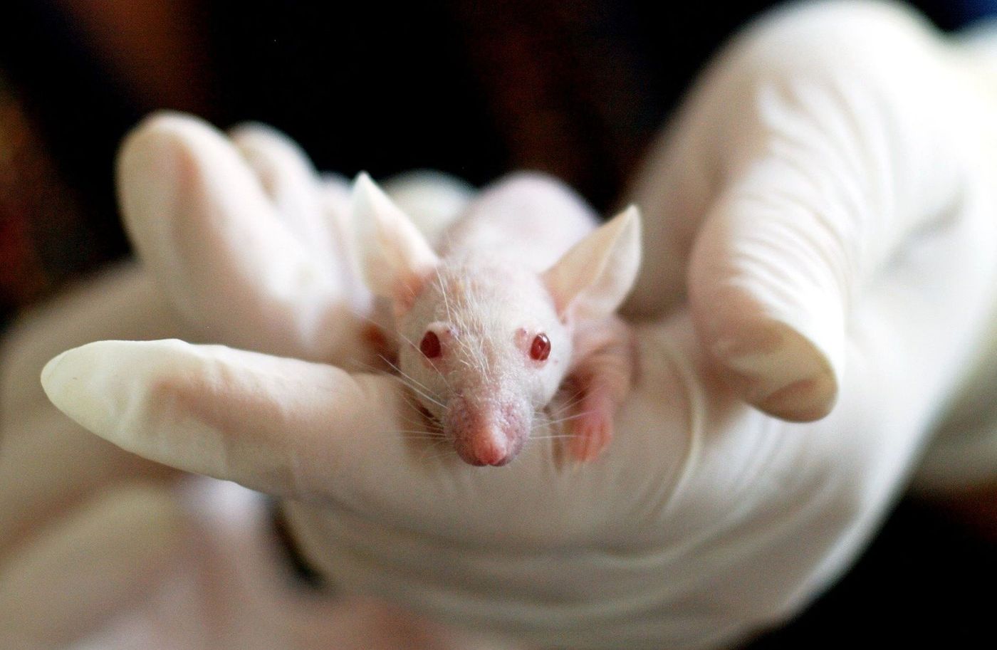
One challenge of working with mouse cancer models is obtaining precise tumor growth measurements. Researchers often use calipers that manually measure two dimensions of a murine tumor, but this method is open to unintentional user bias. While researchers can perform advanced imaging methods on laboratory mice, such as magnetic resonance imaging (MRI), these techniques only provide low-resolution measurement capability. Further, many advanced tools available for monitoring tumor growth in mice often prove time-consuming and expensive. Importantly, most imaging technologies available for mice require anesthetization which, notably, is typically not necessary when using similar methods in patients.
A new study published in Science Advances has demonstrated a unique approach to continuously monitoring the growth or regression of murine tumors in the micrometer range. This technology, referred to as FAST (flexible autonomous sensors measuring tumor volume regression), wraps around a tumor near the animal's skin and provides researchers with a wealth of essential data.
The flexible electronic sensor conforms to the mouse and detects strain induced by a growing, or shrinking, tumor providing regular measurements to the researchers. The authors describe how this method offers three improvements over standard murine tumor measurements. First, FAST records measurements every five minutes providing a complete picture of tumor growth over time, allowing researchers to know how quickly an investigational treatment works. Such measurement frequency remains highly infeasible by any standard methods of monitoring murine tumor growth. Second, the authors explain that the sensor improves measurement accuracy by distinguishing changes in tumor size too small to detect with standard methods. Third, FAST is autonomous and noninvasive, further enhancing the ability to achieve an accurate picture of tumor regression while subjecting the animal to minimal stress or discomfort.
The authors conclude that the ability of FAST to continuously, autonomously, and accurately monitor changes in tumor volume makes it an excellent candidate to replace current tumor measurement methods. This technology can significantly improve pre-clinical research strategies by providing high-throughput screening of novel drugs under development. This technology may expedite the generation of new drugs that will benefit cancer patients and extend survival.
Sources: Dis Model Mech, Sci Transl Med, Sci Advances