SARS-CoV-2 infections, which cause COVID-19, can wreak havoc on the body. Early on in the pandemic, doctors noted that while the disease began as a respiratory infection, many more organ systems were often affected. It's been suggested that the virus may invade tissues like the liver or kidneys directly. In other cases, the definitive presence of the virus in cells was less clear, but inflammation caused by the viral infection was taking an obvious toll on organs like the heart and brain.
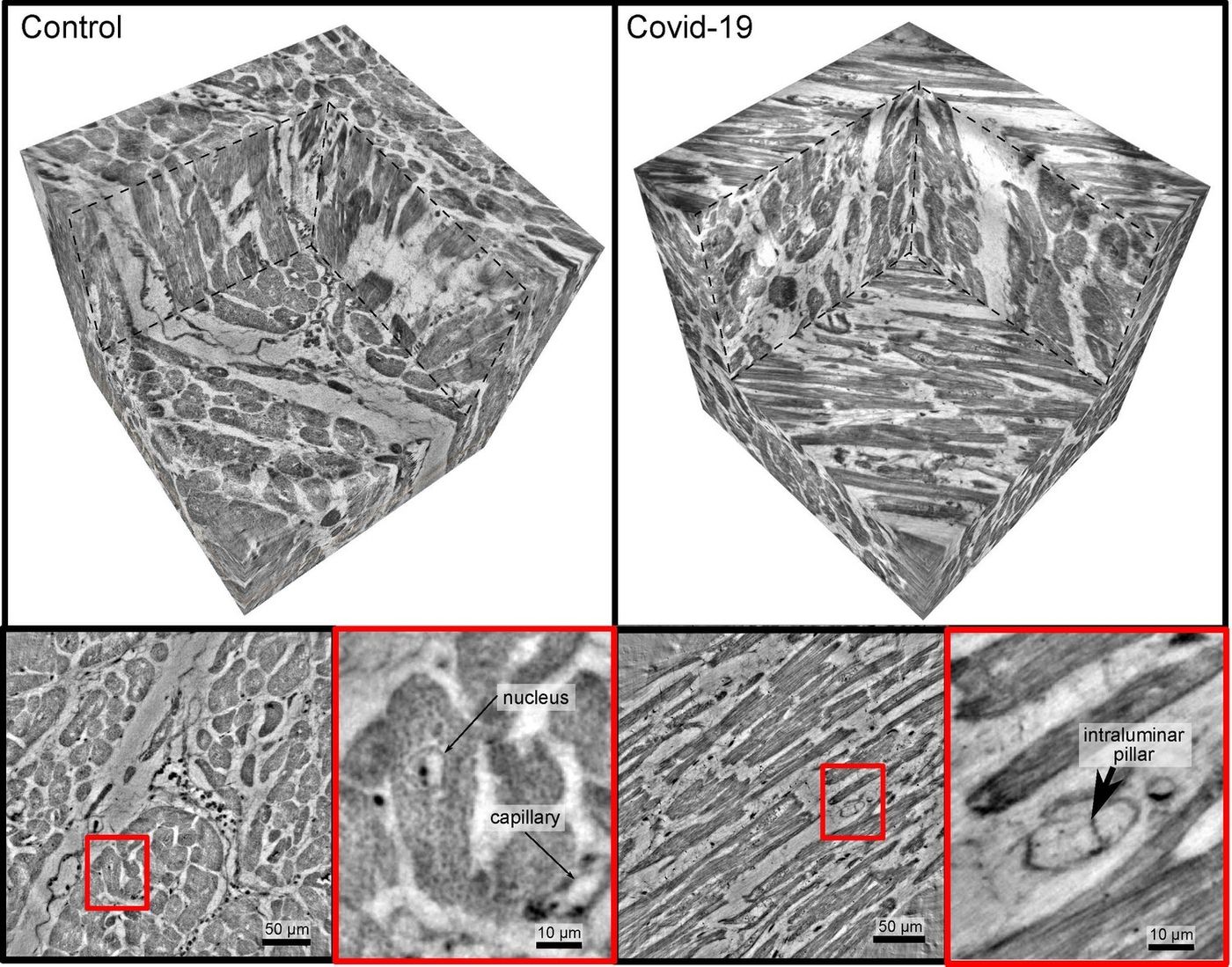
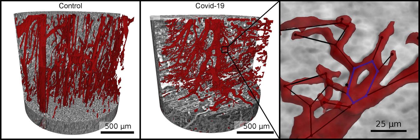
Scientists have now shown that in people who died from COVID-19, there were significant changes in their hearts. The researchers used an imaging tool called synchrotron radiation, which is an intensely bright X-ray in this study. It can analyze tissue at high resolution in three dimensions to reveal damage at the microscopic level.
This work showed that in patients with COVID-19, the capillary networks that flow through the heart had been chaotically disordered, and new vessels had formed and split away, compared to healthy heart tissue. The findings show how the formation of new vessels, a phenomenon known as intussusceptive angiogenes, can drive apthologies associated with COVID-19, and have been reported in eLife.
The study authors relied on machine learning to process the network of capillaries, so they had to manually label data to start. In doing so, they compared the tissue architecture, said first study author Marius Reichardt of the University of Göttingen. This indicated that the tissue affected by COVID-19 was very different from healthy tissue, or even tissue that came from individuals with diseases like myocarditis or severe influenza, study co-leaders Professor Tim Salditt of the University of Göttingen and Professor Danny Jonigk of Hannover Medical School (MHH).
The researchers were able to demonstrate that any clinic with a small X-ray source could perform diagnostics the way they did; clinicians do not need to obtain the level of detail acquired in the research to get useful information. The study authors are interested in broadening this method so normal tissue patterns can be identified with X-ray imaging and machine learning, then used to create diagnostic tools.
Experienced research scientist and technical expert with authorships on over 30 peer-reviewed publications, traveler to over 70 countries, published photographer and internationally-exhibited painter, volunteer trained in disaster-response, CPR and DV counseling.