Many of the things biologists want to study are extremely small, and researchers now have many powerful tools for getting a magnified view of a specimen. But several years ago, scientists decided to take another approach and make the sample under study larger; the tool they created was called expansion microscopy (ExM).
Since being developed, ExM has been used to image cells and their contents with incredible spatial resolution. In the process, a specimen is embedded into a polymer that crosslinks the proteins, and then water is used to greatly expand everything. In this way, researchers can get a better look at the structures of the embedded proteins. Now, researchers have shown that it can be used for more than that.
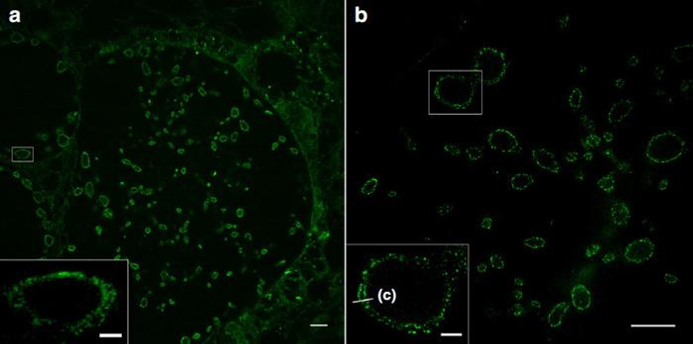
"This method was previously limited to proteins. In the journal Nature Communications we are now presenting a way of expanding lipids and thus cell membranes," said senior study author Professor Markus Sauer of the Biocentre of Julius-Maximilians-Universität (JMU) Würzburg in Bavaria, Germany.
Cell membranes are primarily made of lipids, fatty molecules that are arranged in a very specific way. Molecules called sphingolipids are one part of these membranes. Researchers in the Seibel lab have generated synthetic sphingolipids that can be added to cell cultures, which then get incorporated into the membranes of the cells in culture. That enables scientists to mark the membranes, and then expand them using a swellable polymer, as in ExM.
With this technique, the researchers demonstrated that ExM, together with structured illumination microscopy (SIM) can be used to view different cell membranes and how they interact with proteins, at a resolution of ten to twenty nanometers. This will provide greater insight into the membranes of cells and their nuclei as well as the membranes of cellular organelles.
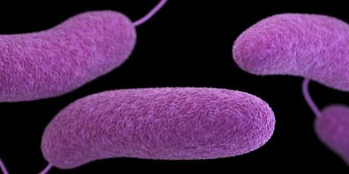
These sphingolipids can also be easily incorporated into bacterial membranes. Thus, the membranes of pathogenic bacteria can be visualized with a tool other than electron microscopy, which may help us learn more about penetrating these membranes - and fighting the pathogens.
"With the new super-resolution microscopic methods, we now want to investigate bacterial infection mechanisms and causes of antibiotic resistance. What we learn in the process could possibly be used for improved therapies," said study co-author Professor Thomas Rudel.
It may also be possible to add these sphingolipids to viral membranes, but more work will be needed to know for sure.
Experienced research scientist and technical expert with authorships on over 30 peer-reviewed publications, traveler to over 70 countries, published photographer and internationally-exhibited painter, volunteer trained in disaster-response, CPR and DV counseling.