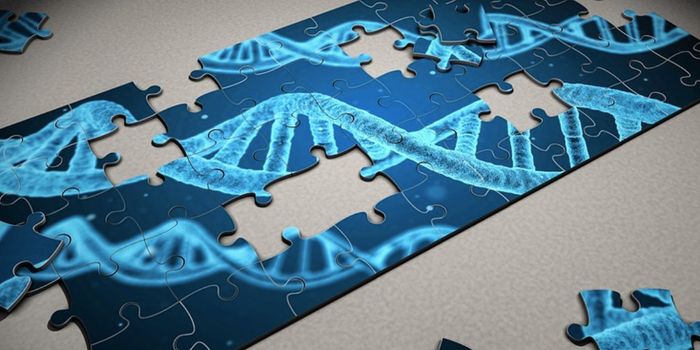
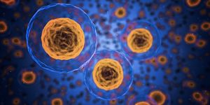
Many types of cells have to be able to move around, such as during the development of the body, or when immune cells have to reach the site of an infection or injury. To do so, cells may employ lamellipodia, thin extensions that can extend, attach to a surface, and pull the rest of the cell forward. A dense network of proteins called actin filaments forms the cell's cytoskeleton and is also found within these lamellipodia. A group of seven proteins known as the Arp2/3 complex uses existing actin filaments to generate new ones, generating the branching forces that can take the cell to new places.
Existing methods enabled scientists to study the Arp2/3 complex's structure is one of two ways: either in isolation when the structure was inactive, or once bound to actin filaments. In the first method, the inactive conformation won't reveal much to researchers about how the complex works to sprout new filaments. The second method uses electron tomography, which shows the complex in action but the resolution is not great.
"Previous electron tomography data of Arp2/3 complexes bound to actin filaments in a test-tube environment was too imprecise, making it impossible to unambiguously tell where the individual elements of the complex must be located," explained Florian Fäßler, a postdoctoral researcher in the lab of IST Austria professor Florian Schur.
In new research reported in Nature Communications, scientists in the Schur lab have imaged many Arp2/3 complexes inside of mouse cells' lammelipodia while they were in action. The researchers used powerful cryo-electron microscopy tools to rapidly freeze the cells in place, then image them from many angles. This generated enough data to reconstruct three-dimensional images of 10,000 different, active Arp2/3 complexes.
"We said to ourselves: Okay, we are going into the cell, where the environment is much more intricate because there is not only the protein complex and actin filaments but all sorts of other things as well. But this was the only way we were able to maintain this network in such a way that we could determine its structure," said molecular biologist Florian Schur.
This study provides unprecedented insight into the relationship between Arp2/3 complexes and actin filaments. Now researchers can learn more about its function and how it may play a role in disease.
"What we have done is to go as far as is currently possible with such complex samples in terms of methodology and resolution. With the current resolution, we have gained new biological insights, but it was also a methodological advance to show: It is possible," Schur noted. "We are just starting to realize the full potential of cryo-electron tomography."
Experienced research scientist and technical expert with authorships on over 30 peer-reviewed publications, traveler to over 70 countries, published photographer and internationally-exhibited painter, volunteer trained in disaster-response, CPR and DV counseling.