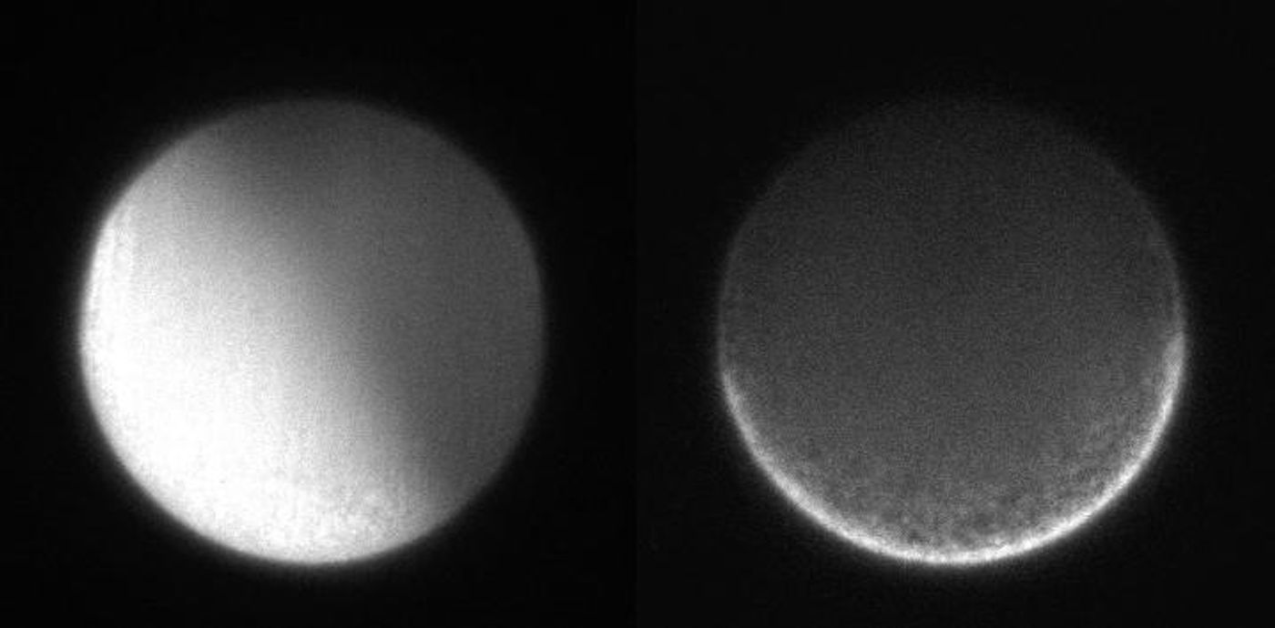
Animals grow from what looks like a clump of cells, but those cells organize into specific patterns, laying the right foundation so that head, tail, back, and belly all end up in the right place, facing the right direction. Now researchers are getting closer to understanding precisely how the body axis is formed. In new work reported in Molecular Biology of the Cell, scientists captured images of the beginning of development. Their work, which used a sea squirt model, has suggested that both parents can influence the orientation of their offspring's body. In the sea squirt, the back-to-belly axis is set by the mother while the head-to-tail axis is set by the father.
"The most interesting and mysterious part of developmental biology is the origin of the body axis in animals," said researcher Tomomi Tani, who worked as a Marine Biological Laboratory (MBL) scientist at the time of the research, and is now with Japan's National Institute of Advanced Industrial Science and Technology.
"Both the maternal and the paternal cues are required to establish the body plan of the developing animal embryo," Tani added.
It's been thought that in the egg, filaments made of a protein called actin help set the body axis by propelling the rearrangement of stuff in the cell cytoplasm after it's been fertilized. Observing this process has been difficult, however, because it happens fast in a very small space. In this work, Tani and Hirokazu Ishii used a tool called a fluorescence polarization microscope to overcome these hurdles.
"Using polarized light for looking at dynamics of molecular order is a tradition of MBL imaging," Tani noted.
Polarized light waves only oscillate in only one direction or partially, therefore a filter lets polarized light pass through in one orientation but blocks it when rotated. Fluorescent probes were used to illuminate actin in sea squirt eggs, and the rigid connection between the probe and actin enabled the researchers to detect actin's orientation when illuminated by polarized light.
The researchers could tell when all of the actin filaments were oriented in one direction, or if they were pointed in various directions that could be detected as well. The scientists saw that in unfertilized eggs, the actin was oriented randomly. But after being fertilized, a wave of calcium ions moved through the eggs and the actin filaments lined up in one direction, contracting at a right angle to the upcoming back-to-belly axis, and moving the cytoplasm.
The researchers are planning to continue this work to understand more about the process of embryonic development and how it might go awry.
"We hope that the molecular orders in the cytoskeleton tell us something like 'field lines' of mechanical forces that organize the morphology of multicellular organisms," Tani said.
Experienced research scientist and technical expert with authorships on over 30 peer-reviewed publications, traveler to over 70 countries, published photographer and internationally-exhibited painter, volunteer trained in disaster-response, CPR and DV counseling.