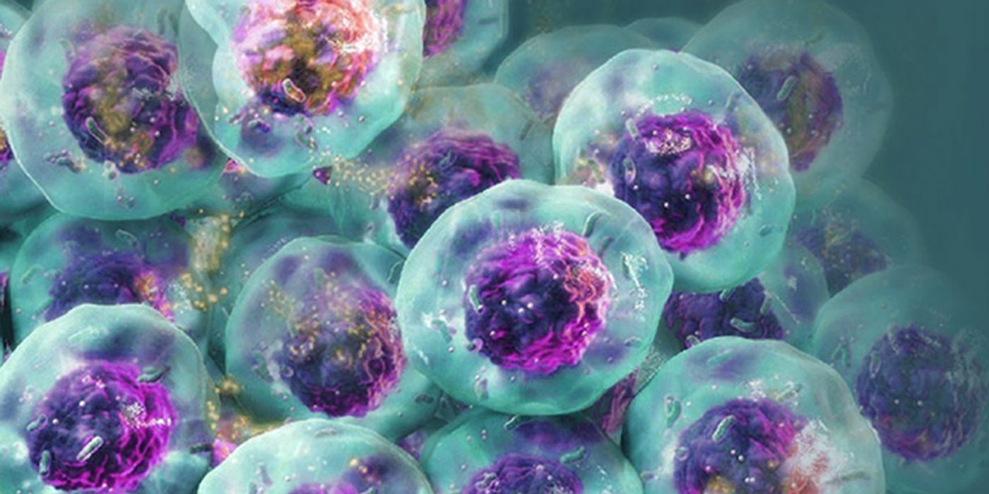
Researchers are increasingly relying on cells grown in three-dimensional (3D) structures to help answer their research questions. Monolayer, or 2D cell culture, was the go-to cell culture method for the past century. Now, the need to better represent in vivo conditions is driving the adoption of 3D cell culture models. Cells grown in 3D structures better mimic tissue-like structures, better exhibit differentiated cellular functions, and better predict in vivo responses to drug treatment.
Switching to 3D cell culture models comes with challenges. Methods to interrogate these models need to be adaptable and reliable for the many types of 3D models. Some of the most popular 3D models include spheroids grown in ultra-low attachment plates and cells grown in an extracellular matrix, such as Matrigel® from Corning. Even more complex models include medium flow over the cells in microfluidic or organ-on-a-chip devices. Will an assay originally developed for cells grown in monolayer perform consistently with various 3D models? How is measuring a cellular marker different when cells are grown in 3D models compared to monolayer growth?
One such marker that researchers want to interrogate with 3D models is caspase 3 activity. Caspase 3 activity is a marker of apoptosis or programmed cell death. If a drug treatment activates caspase 3 and induces apoptosis, it could potentially be an effective cancer drug. Because 3D cell culture better mimics tumor cell growth, cancer researchers are increasingly testing potential drugs that activate caspase 3 with 3D models.
The Caspase-Glo® 3/7 3D Assay was recently introduced to help researchers detect caspase 3 activation in 3D models. The new 3D-labeled version of the assay is based on chemistry originally developed for monolayer cell cultures. To ensure the new assay generates reliable results in multiple 3D models, a panel of cells was grown in both spheroids and Matrigel®-embedded tissues. Parameters including lysis of spheroids, signal linearity, and dose-response relationship were evaluated to ensure the assay reagent could be used with a wide range of 3D models.
In addition, 3D spheroids of varying sizes were treated with drugs to see how the size difference would influence the apoptotic response. Results showed that the apoptotic signal in liver cancer cell spheroids (HepG2) induced by Panobinostat (an approved chemotherapeutic drug) was proportional to spheroid size. The larger spheroids were more susceptible to the drug treatment, indicating that the size of the spheroid impacted how cells responded to the drug. The data reinforce the advantage of switching to 3D models, which better mimic how cells or tumors grow in our body and is a better model for researchers studying cancer treatments.
Interested in learning more? See more data in this scientific poster: Caspase-Glo® 3/7 3D: Validation for Spheroids and Matrigel® Embedded Tissues or explore more resources about 3D cell culture in Promega’s 3D Cell Culture Guide.