Since Shinya Yamanaka developed a method to create stem cells from regular adult skin cells using genetic reprogramming techniques (the resulting cells are known as human induced pluripotent stem cells or iPSCs), research in the field has accelerated rapidly. Stem cells have been used to create miniature models of human organs that are made up of different cell types that typically self-assemble into a three-dimensional structure, called organoids. These cultured models are becoming more and more complex. There has even been discussion about whether organoids that are meant to mimic brains will gain some form consciousness and the ethical implications of such research.
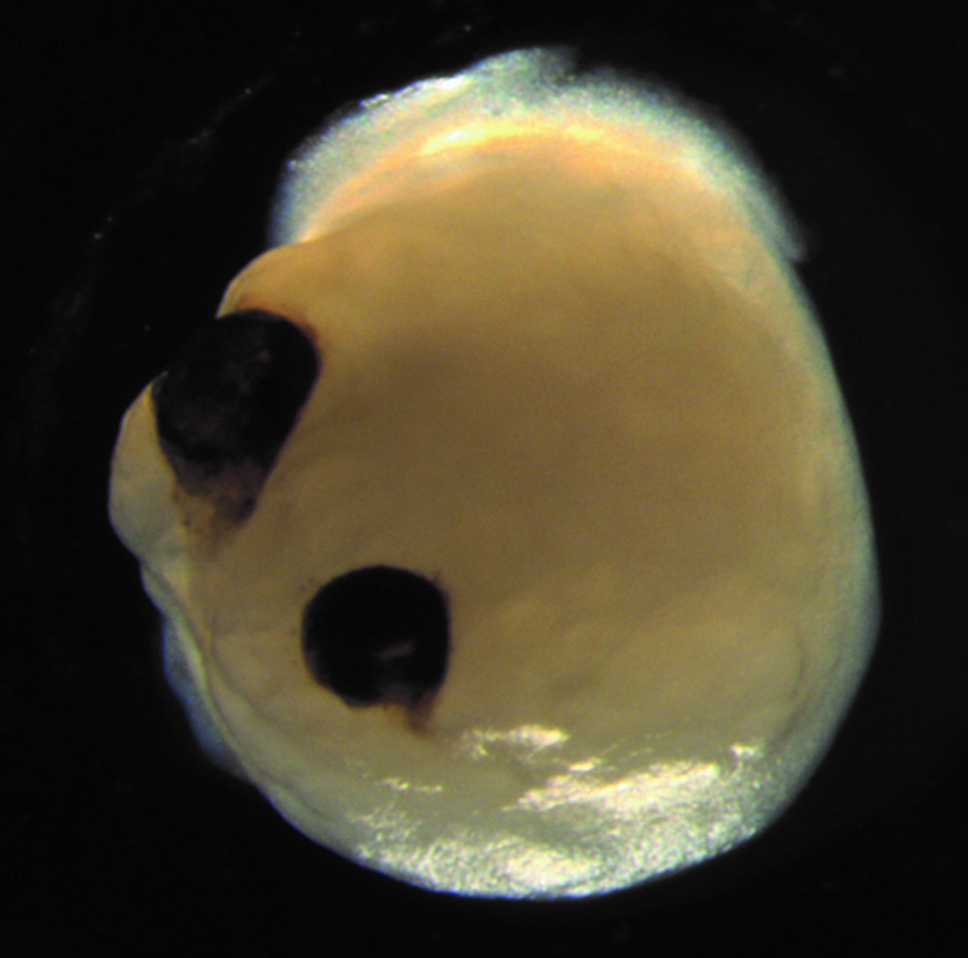
Now researchers have reported in Cell Stem Cell that they created brain organoids that went on to develop a structure that makes up part of the eye where the retina links to the optic nerve, called the optic cup. These bilateral and symmetrical features spontaneously grew, which suggests that the intrinsic abilities of certain groups of cells are quite robust.
“Our work highlights the remarkable ability of brain organoids to generate primitive sensory structures that are light sensitive and harbor cell types similar to those found in the body,” said senior study author Jay Gopalakrishnan of University Hospital Düsseldorf. “These organoids can help to study brain-eye interactions during embryo development, model congenital retinal disorders, and generate patient-specific retinal cell types for personalized drug testing and transplantation therapies.”
Brain organoids have now been generated from iPSCs and used in many studies to investigate development and disease. Scientists have shown that iPSCs can be utilized to engineer a structure that is similar to the optic cup. Human embryonic stem cells have also been used to produce an optic cup. This previous research was mainly focused on the retina, however, and was not part of a brain organoid.
In this study, the researchers modified a method that creates neural tissue from iPSCs. The optic cups arose after about thirty days or more, maturing within fifty days. This time course is thought to be similar to the progress of retinal development in a human embryo.
The scientists used four iPSC donors to make 314 brain organoids, and 72 percent of them went on to grow optic cups. Various cell types that are found in the retina were present in the organoids, and they carried neuronal networks that were capable of responding to light. The optic cups had some connections to different portions of the brain organoids, and generated some corneal and lens tissue.
“In the mammalian brain, nerve fibers of retinal ganglion cells reach out to connect with their brain targets, an aspect that has never before been shown in an in vitro system,” noted Gopalakrishnan.
The researchers want to determine how to maintain the optic cups for a longer period so they can be used to study retinal disorders.
Experienced research scientist and technical expert with authorships on over 30 peer-reviewed publications, traveler to over 70 countries, published photographer and internationally-exhibited painter, volunteer trained in disaster-response, CPR and DV counseling.