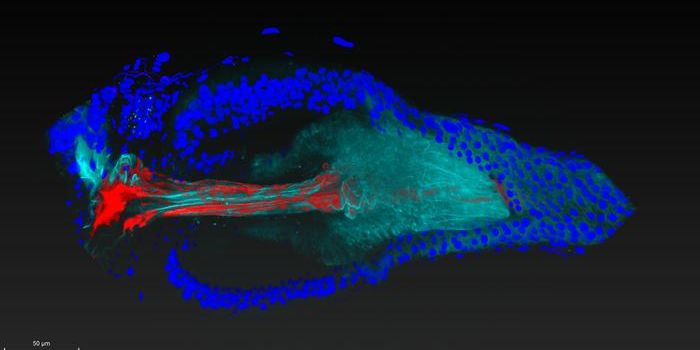
Since the first cells were observed under a microscope, scientists have been finding new ways to illuminate their contents. The more details we can reveal, the more we'll know about how cells normally work, and what happens when their functions go wrong. A wide array of microscopy techniques have been developed in recent years. Scientists have now combined two methods to create a technique for viewing the action inside and outside of live cells at an incredible level of detail. Two reports outlining this technique have been reported, one earlier this year in Nature Communications, and another in ACS Nano.
“The methods currently available present many technical challenges to observe live cells at such a granular level,” said Georg Fantner, the head of EPFL’s Laboratory for Bio- and Nano- Instrumentation (LBNI). “Techniques such as electron microscopy allows unmatched resolution of the cell surface at the nanoscale, but it requires placing samples under vacuum and bombarding them with electrons." The technique is too harsh, and live organisms aren't able to survive the treatment. "Another common method is fluorescence microscopy. Although it lets you observe samples without destroying them, to have sufficient resolution to resolve the three dimensional surface of the cell is difficult. Additionally, the dose of photons required can [cause] cell damage.”
In this work, the researchers used a tool called stochastic optical fluctuation imaging (SOFI) that can be applied to cells to see specific molecules or processes, and combined it with a technique called scanning ion conductance microscopy (SICM). In SICM, cells are usually touched with a probe that maps the cell surface, revealing its topography. The native state of live cells won't remain intact when the cell comes in contact with the probe, however. So the investigators created a new type of probe from a glass nanopore, which can use ion flow to sense the cell surface without making contact.
Scientists can use this technique to look at both the exterior and interior of cells simultaneously, seeing what's happening inside while also getting a topographic map of the surface.
This new technology could help scientists learn more about how cells migrate, differentiate, or communicate, suggested the researchers. Cells often have to detect and react to their environment, and this tool can help us understand the mechanisms underlying those processes.
Experienced research scientist and technical expert with authorships on over 30 peer-reviewed publications, traveler to over 70 countries, published photographer and internationally-exhibited painter, volunteer trained in disaster-response, CPR and DV counseling.