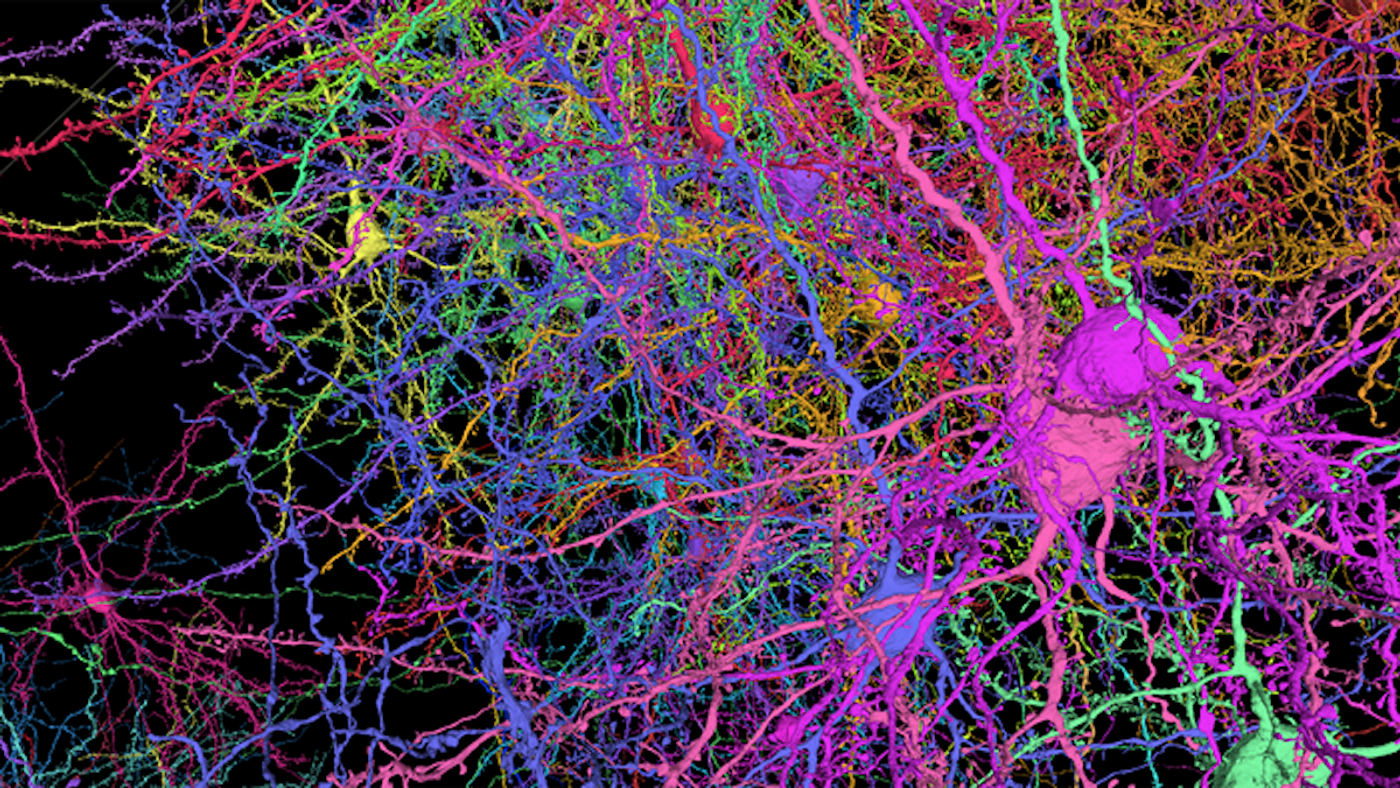
A detailed map of thousands of neurons in the mouse brain was created by a team of researchers from the Allen Institute, Baylor College of Medicine, and Princeton University. This wiring diagram contains 200,000 brain cells and nearly 500 million synapses that sit in a chunk of the visual cortex of the mouse brain that's about the size of one grain of sand. This map has an unprecedented level of detail that can be viewed in three dimensions, and it's publicly available. Like other recent diagrams that have been created from the fruit fly and human brains, this one was generated with electron microscopy.
While this research was intended to provide important insights into signaling in the brain, it also improved machine learning techniques. Various cell types and structures can be seen, including neurons, astrocytes, and blood vessels, as well as the internal parts of some cells.
“We’re basically treating a brain circuit as a computer, and we asked three questions: What does it do? How is it wired up? What is the program?” said R. Clay Reid, M.D., Ph.D., a Senior Investigator at the Allen Institute. “Experiments were done to literally see the neurons’ activity, to watch them compute.” Researchers will be able to view the neural circuitry and follow the wiring. Eventually, Reid added, we may be able to read the program of the brain.
At the start of the research, a team at Baylor recorded the activity in the visual cortex of mice as they viewed images, movies, or natural sciences. Next, scientists at the Allen Institute dissected that portion of the mouse brains, creating thin slices of the visual cortex and imaging them in fine detail with electron microscopes; 150 million images were generated from this effort. At Princeton, researchers used machine learning to decipher the identity of each cell and cellular component. This work created intricate data on cellular connections and cells that extend across the brain section.
The research took about five years to complete. “Today, we have been rewarded by breathtaking new vistas of the mammalian cortex,” said H. Sebastian Seung, Ph.D., Professor of Neuroscience and Computer Science at Princeton University. “As we transition to a new phase of discovery, we are building a community of researchers to use the data in new ways. Only the collective efforts of this community can realize the resource’s potential for enabling new discoveries about the brain.”
Experienced research scientist and technical expert with authorships on over 30 peer-reviewed publications, traveler to over 70 countries, published photographer and internationally-exhibited painter, volunteer trained in disaster-response, CPR and DV counseling.