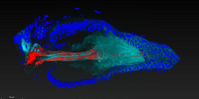
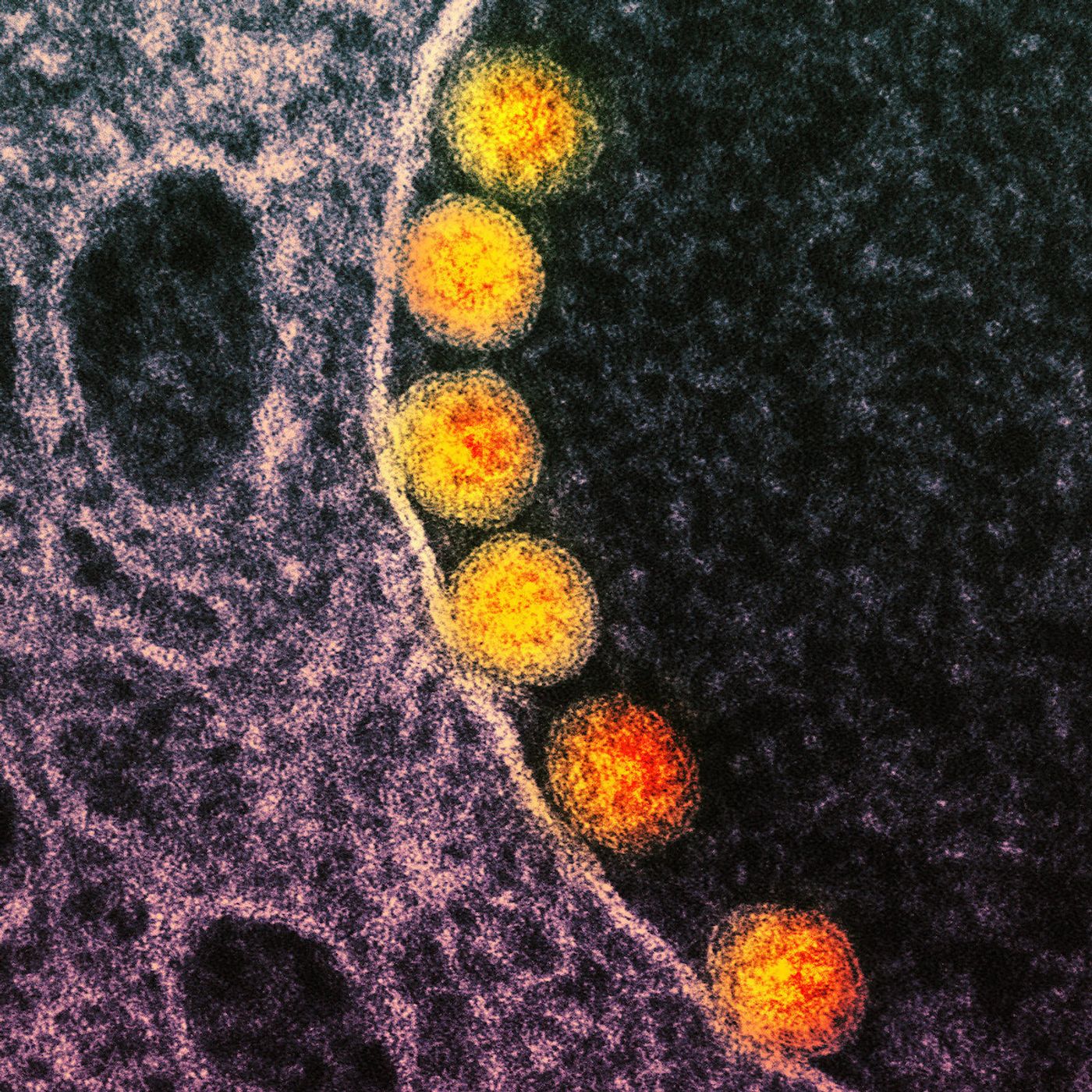
Scientists have caught a virus in the act of infecting a cell, and captured images of the action in real-time and three dimensions. This work was made possible by combining a technique called lattice light sheet microscopy with chemical and genetic tools that labeled various molecules so they could be visualized.
In the video, a virus that was genetically engineered to generate the SARS-CoV-2 spike protein, which is seen in pink, moves to the surface of a cell. The viral protein is then taken up by a cellular sac called an endosome. The virus fuses with the membrane of the endosome, which allows the virus to then release its genetic material, seen in blue, into the cell, which is then infected. That process triggers a cycle in which the cell becomes a host for the virus, and more viral particles are produced with the cell's machinery.
In the second part of the video, many viral particles can be seen inside the cell. A snapshot was taken every four seconds over four minutes to create this video. The work has been reported in the Proceedings of the National Proceedings of Sciences (PNAS).
This video has shown that the SARS-CoV-2 virus, which causes COVID-19, cannot fuse with a cellular membrane to release its contents unless the virus and host cell are in a slightly acidic environment, with a pH between 6.2 and 6.8. This pH is comparable to many body fluids including saliva.
Endosomes are also slightly acidic themselves; enzymes in the endosome or on the surface of cells need that acidic environment too. TMPRSS2, a human enzyme that is known to aid SARS-CoV-2 infection, can cut the spike protein and encourage membrane fusion and the infection of the cell.
The human nose, where SARS-CoV-2 tends to infect cells, also has an acidic environment.
"Amusingly enough, measuring the pH of the nostril cavity has rarely been done before," commented co-senior study author Tomas Kirchhausen, a professor at the Blavatnik Institute and Boston Children's Hospital.
This research could provide new insights into how to prevent SARS-CoV-2 infection, and could apply to other viruses as well.
Sources: Harvard Medical School, Proceedings of the National Academy of Sciences