Scientists have developed an advanced imaging technique for studying mitochondria. These organelles are often known as the powerhouses of the cell because they generate energy, but studies have shown there is a lot more to mitochondria than just energy production. They have roles in cell division and helping a cell survive stressful times. Mitochondria also carry their own genomes and can even produce their own proteins with their own ribosomes. Dysfunctions in mitochondria can lead to a host of disorders; there are mitochondrial diseases that occur when mutations arise in mitochondrial DNA, and mitochondria have also been linked to some types of cancer and neurodegenerative diseases.
This new imaging technology will allow scientists to observe detailed parts of mitochondrial structure and quantify very subtle changes that happen inside of mitochondria, and how they may relate to events in the cell. The work has been reported in the Journal of Cell Biology.
“We now have a powerful new toolkit for detecting and quantifying structural, and thus functional, differences in mitochondria, for example, in diseased versus healthy states,” said senior study author Danielle Grotjahn, PhD, an assistant professor at Scripps Research Institute.
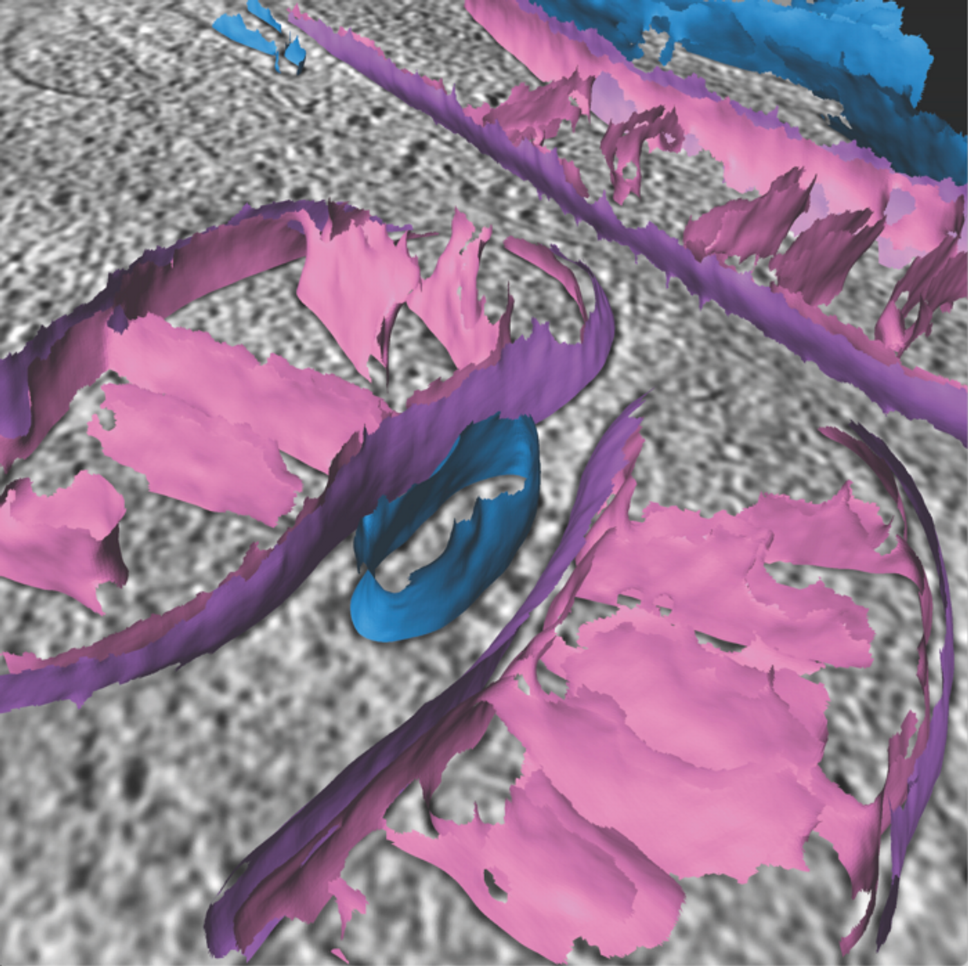
Mitochondria have both an outer membrane and an inner membrane that contain molecules that promote crucial biochemical reactions. The morphology or appearance of structures in the mitochondria can change significantly based on mitochondrial function or whether a cell is under stress. The structural changes can work as markers of conditions in the cell, and with this tool, researchers will be able to detect and quantify them.
The technique utilizes computational tools to assess images generated by cryo-electron tomography (cryo-ET), which uses electrons instead of light to image biological samples in three-dimensions. The investigators called this a “surface morphometrics toolkit.” It maps and measures bends in the inner mitochondrial membrane, the gaps between the membranes, and other structural elements that might serve as markers of various conditions.
The researchers also used the surface morphometrics toolkit to study mitochondria in cells that are experiencing endoplasmic reticulum stress, which is sometimes seen in neurodegenerative diseases. This revealed drastic alterations in structures like the inner membrane, and the gap between membranes.
The researchers also noted that this method is not limited to mitochondria, and can be used to study other cellular organelles.
Experienced research scientist and technical expert with authorships on over 30 peer-reviewed publications, traveler to over 70 countries, published photographer and internationally-exhibited painter, volunteer trained in disaster-response, CPR and DV counseling.