
![[Guide] 7 Strategies to Boost Laboratory Collaboration](https://d3bkbkx82g74b8.cloudfront.net/eyJidWNrZXQiOiJsYWJyb290cy1pbWFnZXMiLCJrZXkiOiJjb250ZW50X2FydGljbGVfcHJvZmlsZV9pbWFnZV83YzBjZWIwM2Y5YzI4MmFlYzBhZDZhMTcyNTQ1ZGU3YmE4Y2MzMDYyXzUxNDkuanBnIiwiZWRpdHMiOnsidG9Gb3JtYXQiOiJqcGciLCJyZXNpemUiOnsid2lkdGgiOjcwMCwiaGVpZ2h0IjozNTAsImZpdCI6ImNvdmVyIiwicG9zaXRpb24iOiJjZW50ZXIiLCJiYWNrZ3JvdW5kIjoiI2ZmZiJ9LCJmbGF0dGVuIjp7ImJhY2tncm91bmQiOiIjZmZmIn19fQ==)

Infographics
The Science of Sourdough
Interested in health technology and innovation.
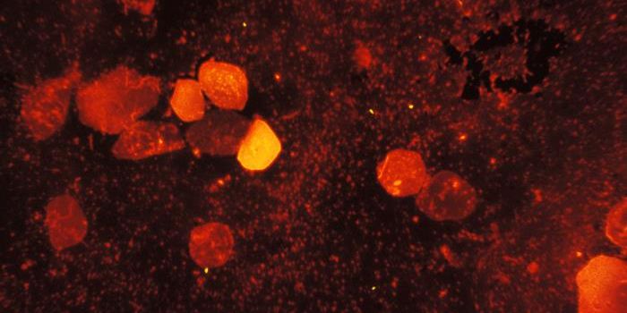
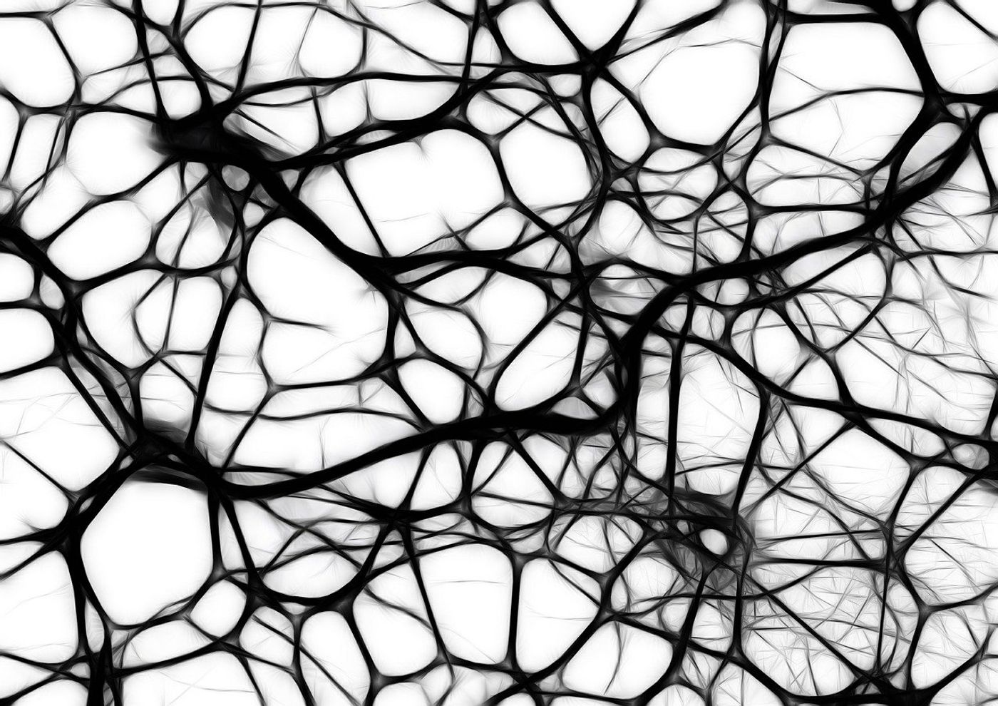
Parkinson’s disease is a devastating condition whereby neurons in a specific region of the brain that controls movement lose functionality or die. This brain region is known as the substantia nigra — which is Latin for "black substance", due to the fact that it contains a high concentration of neuromelanin-containing dopaminergic neurons. As a consequence, those living with Parkinson’s suffer from a suite of debilitating symptoms ranging from shaking, stiffness, and difficulty moving to chronic fatigue and memory difficulties. The early signs of the condition can be incredibly subtle and hard to spot. So, despite being the subject of intense research, there is still neither a definitive diagnostic test nor a cure for Parkinson’s.
Australian Researchers at the University of South Australia are developing a simple neurological imaging tool, which will allow physicians to take a closer look at the specific areas of the brain that are most impacted by Parkinson’s. Brain scans such as PET and SPECT are standard imaging procedures currently used in hospitals. The downside of these imaging tests is, however, the need for patients to be injected with radioactive tracers.
“The significant challenges associated with manufacturing radiotracers and the high cost severely limits patient access to this technology in Australia and elsewhere,” said research lead, Professor Gabrielle Todd.
“Due to the lack of an accurate and accessible diagnostic test for Parkinson’s disease, this results in a high rate of misdiagnosis.”
To complicate matters further, the interpretation of scans can be subjective, heavily influenced by the expertise level of the physician looking at them as well as the quality of the images. This is no easy feat, especially considering the enormous spectrum of variation within the intricate neuroanatomical features between individuals. It’s no surprise that doctors find it a challenge to make the call of whether the substantia nigra of a suspected Parkinson’s patient can be considered “normal”.
Professor Todd and her team are working on an imaging tool that takes the guesswork out of diagnosing Parkinson’s, especially at the early stages. This platform uses MRI scans as an input (a safe, widely available technique that doesn’t require the administration of radioactive substances) with the goal of it being adopted by hospitals everywhere and not just in specialized departments. The research team is close to establishing an easy-to-use method for objectively measuring and evaluating the substantia nigra from MRI images which are compared to a database of normative data.
“Collectively, we hope to make it easier for radiologists to learn about and interpret abnormal substantia nigra MRI findings and to provide neurologists with more certainty about the patient diagnosis,” said Professor Todd.
Sources: University of South Australia, National Institute of Aging.