Genetics & Genomics
Cord Blood Samples Reveal More About the Genetics of Autism
Interested in health technology and innovation.
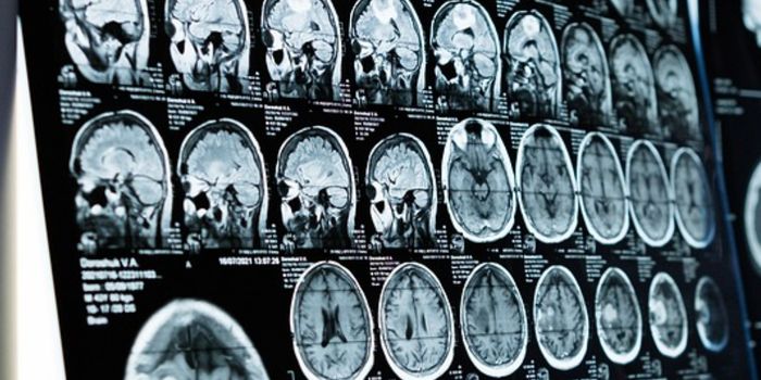
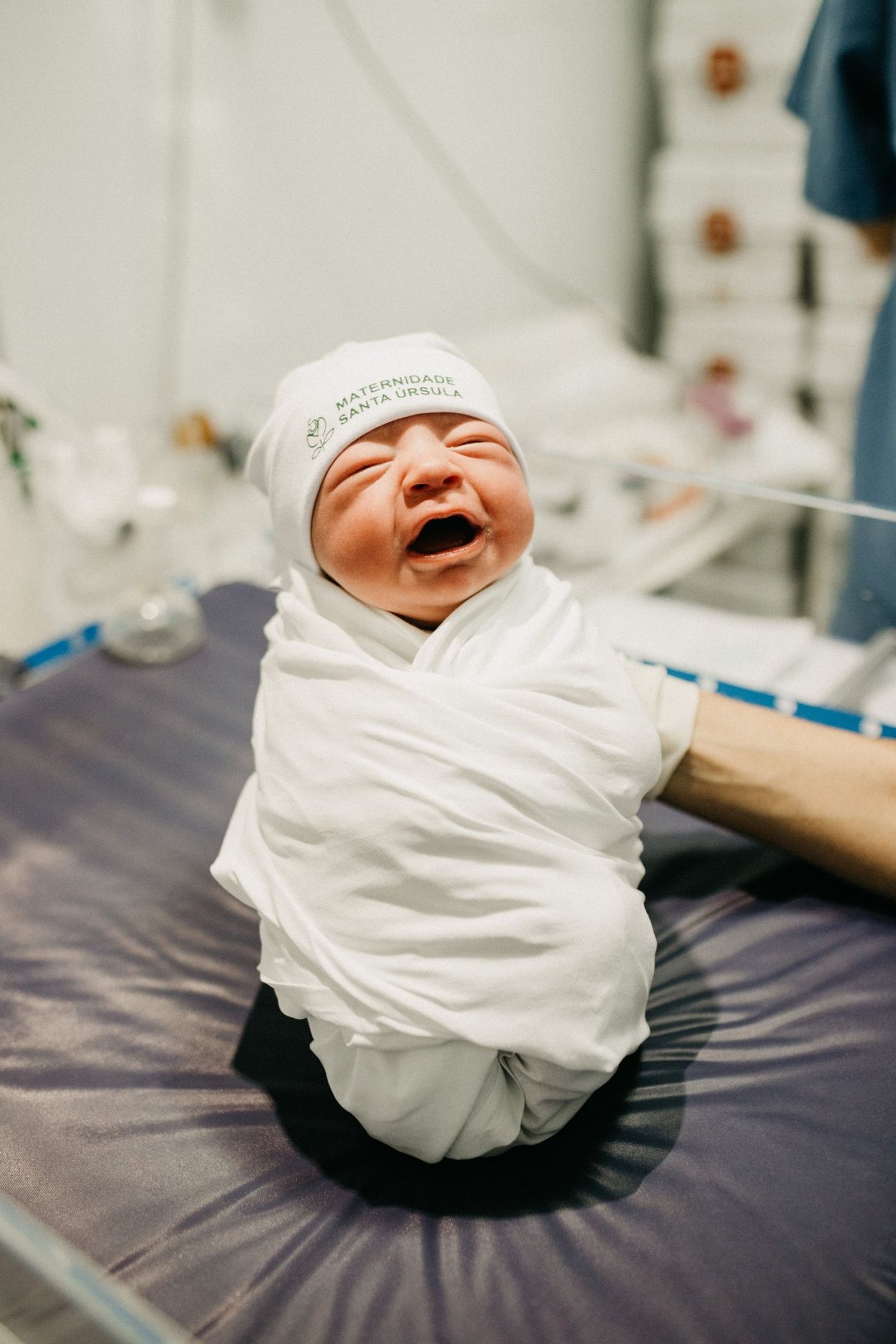
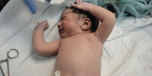
Diagnostic imaging scientists have developed a software tool for predicting the future onset of cerebral palsy in babies born prematurely. Children born earlier than 30 weeks of gestation face a staggeringly elevated risk of problems with motor development. This is in part due to abnormalities in their white matter, deep regions in the brain that contain nerve fibers.
Almost 70 percent of babies born extremely prematurely display these white matter abnormalities and up to 10 percent of them go on to develop cerebral palsy, or CP, a permanent and incurable movement disorder.
According to experts, earlier CP diagnoses lead to earlier interventions, transforming the quality of life for both the children affected and their caregivers. Physical therapy, for example, if initiated early in the child’s development, could help reduce symptoms of CP including stiffness, tremors, and problems with fine motor control. Current neuroimaging technologies are unfortunately not able to achieve this, largely due to sensitivity constraints and highly subjective assessment protocols.
Neonatologist Nehal Parikh from Cincinnati Children’s Hospital Medical Center led a team that developed a computational tool to analyze magnetic resonance imaging (MRI) scans taken of preterm infants, flagging those with signature anatomical defects that correlate with an increased CP risk. This biomarker is termed diffuse white matter abnormality, or DWMA, and can be picked up in MRI scans as regions with more intense signals as compared to surrounding healthy tissues. Neurologists believe that these darker regions are due to inflammation in the white matter.
“Manual tracing of DWMA regions within the brain is highly subjective and produces poor reliability and reproducibility,” said Parikh. “It is clear that we need a better tool for diagnostics. I started working with computer scientists to come up with an algorithm that can segment these lesions.”
The “training” regime for the algorithm involved the team’s computer scientists manually tracing locations of DWMA, normal white matter, gray matter, and cerebrospinal fluid. Once fed with a database of MRI scans from both preterm and normal term babies, the resulting algorithm was found to have an accuracy of over 95 percent.
The next step is integrating automation, for which the team is currently collaborating with MRI vendors to achieve. “Ideally, when the MRI scan is complete, our algorithm would do its job within seconds and provide a value for diffuse white matter abnormality,” said Parikh. “If this value was abnormal, the child would be referred for aggressive early intervention therapies and/or research trials of novel interventions.”
Sources: Scientific Reports, Physics World.