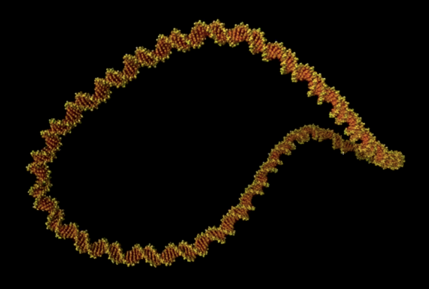
For the first time, researchers have used high-resolution images to generate video footage of DNA as it 'dances' inside a cell, to illustrate the strain and stress that's placed on DNA as it gets compacted into cells. The images are unprecedented in detail, and show the molecule down to the double helix. The scientists added simulations to reveal the position and movement of each atom in the DNA as it twists. The work has been reported in Nature Communications.
There are two meters of DNA in almost every human cell, and it all has to fit inside of the nuclei of these cells. To pack it in, cellular machinery and proteins turn, coil, and loop the DNA molecule, producing twisty loops that are tightly wound and highly dynamic.
In this study, the researchers examined minicircles of DNA, since that's where the molecule gets joined at its two ends into a looped structure, using advanced atomic force microscopy. This allowed the scientists to twist the DNA minicircles even further with computer simulations, and this extra twist made the molecule dance more vigorously.
"The computer simulations and microscopy images agree so well that they boost the resolution of experiments and enable us to track how each atom of the double helix of DNA dances," said study co-author Dr. Agnes Noy, a lecturer in the Department of Physics at the University of York.
The relaxed molecules of DNA didn't do much. But once the scientists applied the extra twist the DNA began to take on very exotic shapes, and it became a lot more dynamic. The resulting 'dance' was found to be important for the DNA to bind to other molecules; the dynamic nature of the DNA allowed it to take on more shapes, giving it more options for biding partners.
“Seeing is believing, but with something as small as DNA, seeing the helical structure of the entire DNA molecule was extremely challenging," said study co-author Dr. Alice Pyne, a Lecturer in Polymers & Soft Matter at the University of Sheffield. "The videos we have developed enable us to observe DNA twisting in a level of detail that has never been seen before.”
Professor Lynn Zechiedrich, a study co-author from Baylor College of Medicine said, “Dr. Pyne and her co-worker’s new AFM structures of our supercoiled minicircles are extremely exciting because they show, with remarkable detail, how wrinkled, bubbled, kinked, denatured, and strangely shaped they are which we hope to be able to control someday.”
“The laws of physics apply just as well to the tiny looped DNA as to sub-atomic particles and galaxies. We can use supercomputers to understand the physics of twisted DNA. This should help researchers such as Professor Zechiedrich design bespoke minicircles for future therapies," noted study supervisor Dr. Sarah Harris, an Associate Professor in the School of Physics and Astronomy at the University of Leeds.
Previous work by scientists at Stanford has indicated that DNA minicircles could be related to aging and may be useful as disease markers.
Experienced research scientist and technical expert with authorships on over 30 peer-reviewed publications, traveler to over 70 countries, published photographer and internationally-exhibited painter, volunteer trained in disaster-response, CPR and DV counseling.