There are 22 bones that compose the human skull. These bones are like plates that join together at flexible joints called sutures. These joints are supposed to allow the skull to expand as the brain develops and gets larger, and then close when growth is complete. But in children born with a condition called craniosynostosis, these sutures close too early and they need corrective surgery. It can be caused by mutations in one gene and though it may be inherited, it's typically random. It's estimated that about one of every 2,200 births is affected.
Researchers have now created a cellular atlas of the developing coronal suture. This work has been reported in Nature Communications. The scientists are hopeful that this study will help create better therapeutic options, or potentially, preventive measures for patients.
"Named for its location at the crown of the head, the coronal suture is affected in a number of birth defects," said lead author D'Juan Farmer, a postdoctoral fellow in the lab of Gage Crump at USC. "These infants and children have to undergo a series of invasive and dangerous surgeries to expand their skulls, and we wanted to understand why the coronal suture is particularly sensitive to gene disruptions. So with an aim toward advancing new interventions for patients, we created the first detailed cell-by-cell description of how this suture develops."
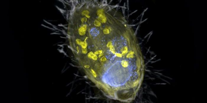
In this study, the researchers used a mouse model to collect cells from the coronal suture as it developed. With thousands of these cells, the researchers assessed protein expression at the single-cell level. This work indicated that there are fourteen different cells types in the developing coronal suture. They also identified several genes that may be directly involved in the production and maintenance of stem cells that generate the bone on either side of the suture.
The research also revealed ligament-like cells that link the skull plates, which stick around into adulthood. Other cell types that sit where the brain and skull bones meet were found as well. The scientists suggested that these may help to regulate the activity of stem cells in the sutures.
In a mouse model of one type of craniosynostosis called Saethre-Chotzen Syndrome, the researchers found that there were fewer stem cells in the suture, and in a more symmetrical distribution than normal mice which had more stem cells in an asymmetrical pattern. The symmetry in the sutures might be causing the skull plate edges to push directly together and fuse, the researchers suggested.
"By examining the very earliest stages of coronal suture development at cellular resolution, our study provides key insights into why this suture is particularly vulnerable to defects in newborns with craniosynostosis," said Farmer.
Additonal research will be needed to confirm these findings in people.
Experienced research scientist and technical expert with authorships on over 30 peer-reviewed publications, traveler to over 70 countries, published photographer and internationally-exhibited painter, volunteer trained in disaster-response, CPR and DV counseling.