With patient cells, researchers created a bioink that was used to 3D print eye tissue. This tissue can now be used as a model for the study of eye diseases. The region of the eye that supports cells that sense light in the retina, the so-called outer blood-retina barrier, can be mimicked with this process. Now researchers have a potentially unlimited supply of tissue that is specialized to individual patients and can be used to investigate disorders like age-related macular degeneration (AMD).
AMD has been shown to begin in the outer blood-retina barrier, said Kapil Bharti, Ph.D., who heads the Section on Ocular and Stem Cell Translational Research at the National Eye Institute (NEI). "However, mechanisms of AMD initiation and progression to advanced dry and wet stages remain poorly understood due to the lack of physiologically relevant human models."
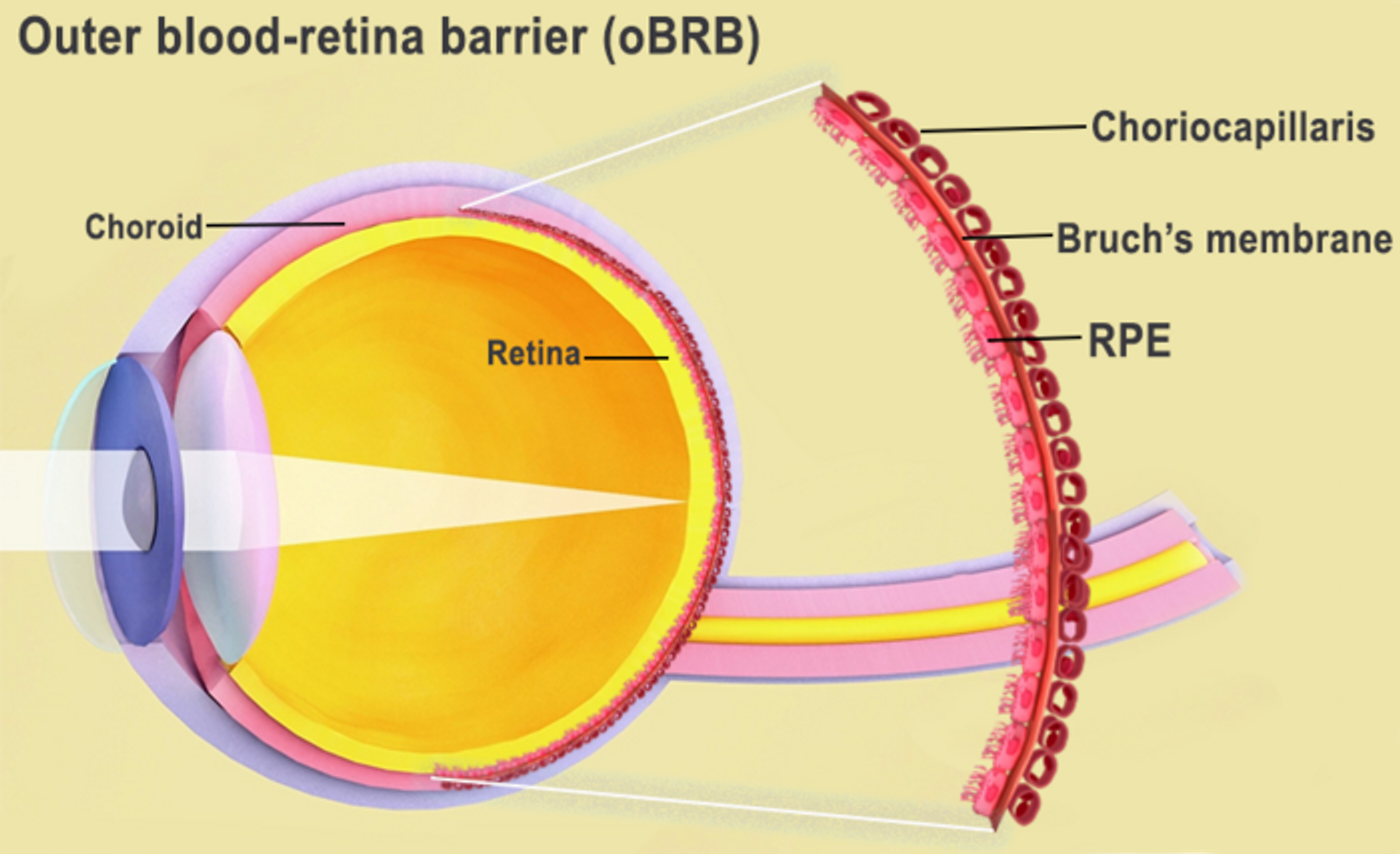
The outer blood-retina barrier is made up of a layer of retinal pigment epithelial (RPE) cells, Bruch’s membrane, and the choriocapillaris, which contains blood vessels. Bruch's membrane regulates nutrient and waste exchange between the RPE and choriocapillaris, but during AMD, lipoprotein deposits known as drusen form and impede its function. The RPE eventually break down, causing the degeneration of photoreceptors and loss of vision.
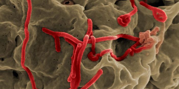
In this work, three types of immature choroidal cells were combined: fibroblasts for structural support, along with pericytes and endothelial cells that compose capillaries, to make a hydrogel. This hydrogel was used as a bioink that was printed onto a scaffold. A dense capillary network formed within days.
After nine days, the opposite side of the scaffold was seeded with RPE cells, and on day 42, it reached full maturity. An analysis of gene expression and tissue functionality indicated that the bioprinted tissue behaves like the outer blood-retina barrier.
When the bioprinted tissue was subjected to stress, it began to take on AMD characteristics, like drusen deposits, then it began to degrade, like what is seen in AMD. Low oxygen levels induced a wet-stage-like appearance in the tissue. When the cells were exposed to anti-VEGF drugs that are used to treat AMD, the tissue became healthy again and vessel overgrowth was halted.
The printed cells facilitates the exchange of cellular signals that are required for normal anatomy in the outer blood-retina barrier anatomy, said Bharti. The presence of RPE cells, for example, caused changes in gene expression in fibroblasts that contribute to Bruch's membrane formation, which was suggested ling ago, but not proven until this model was created.
The researchers are also experimenting with adding new types of cells to the mix to improve the model, and make it even more like normal tissue.
Experienced research scientist and technical expert with authorships on over 30 peer-reviewed publications, traveler to over 70 countries, published photographer and internationally-exhibited painter, volunteer trained in disaster-response, CPR and DV counseling.