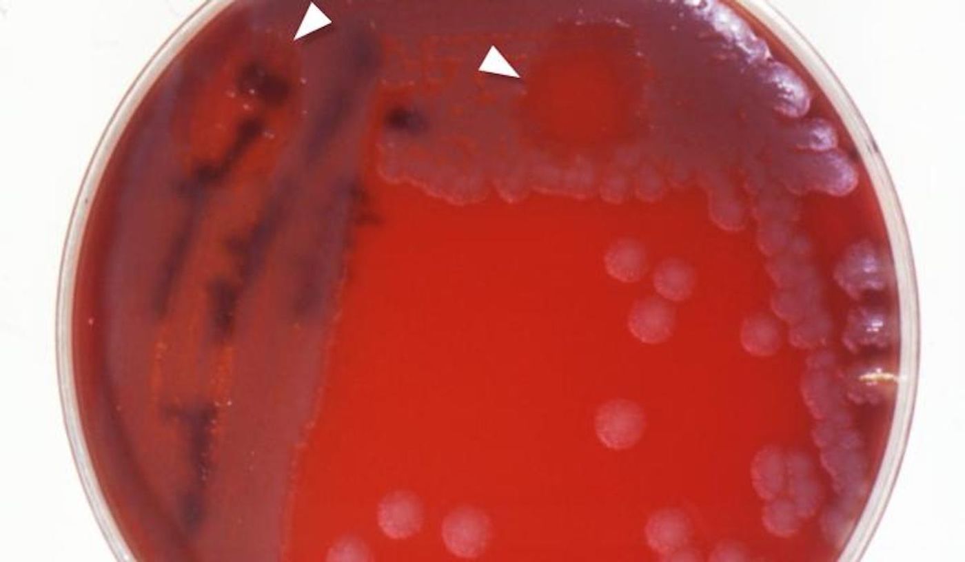
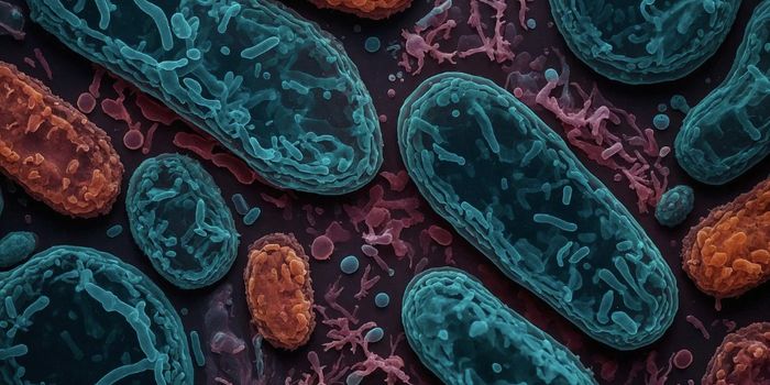
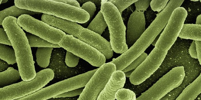
Some viruses only infect bacteria; they care called bacteriophages or phages for short. As antibiotic-resistant bacteria become a greater threat to public health, scientists and clinicians have been looking for creative ways to fight these drug-resistant pathogens, and bacteriophages have already been used to do so in some rare cases. Now scientists want to learn more about phages, which we'll have to do if phages are ever going to be used widely to fight tenacious bacterial pathogens.
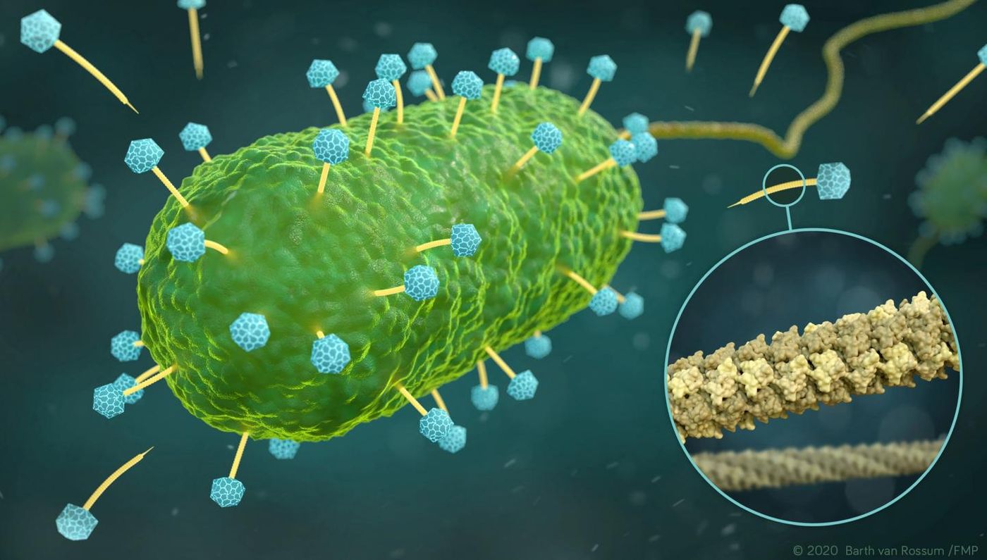
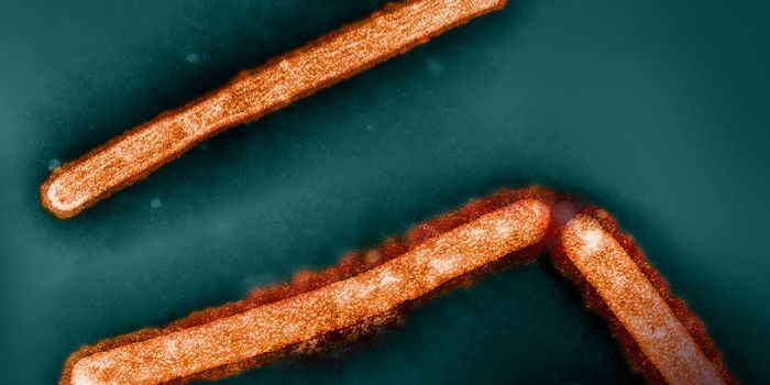
Phages have a structure that enables them to inject their genetic material into host cells. The phage DNA has to move through a tube on the phage as it is released into the host cell. Researchers have now revealed the three-dimensional structure of this critical part of phages, at the atomic level. The work has been reported in Nature Communications.
"The structure and flexibility of the DNA tube attached to the icosahedron-shaped capsid is somewhat reminiscent of a spinal column," noted Professor Adam Lange of Leibniz-Forschungsinstitut für Molekulare Pharmakologie (FMP). "It seems to be perfectly designed for transporting DNA."
In this work, the researchers had to combine two methods to get their results: cryo-electron microscopy and solid-state nuclear magnetic resonance spectroscopy (NMR), as well as computational tools for handling the data. With solid-state NMR, researchers can get a look at flexible structures and small details. Cryo-electron microscopy on the other hand, provides a view of the bigger picture and the overall architecture of the object.
"The key to success was combining the two methods, representing a methodological milestone," commented Lange.
The work revealed six gp17.1 phage proteins that are organized into a stack of rings to form a hollow tube. These rings are linked flexibly together, so the tube is able to bend.
"We are now able to understand how negatively charged DNA is repelled from the likewise negatively charged interior wall of the flexible tube, passing through it smoothly," explained the lead study author Maximilian Zinke. "The bacteria are ultimately destroyed via this pathway."
Researcher Adam Lange is hopeful that this effort will also advance the study of structural biology beyond phage research.
Experienced research scientist and technical expert with authorships on over 30 peer-reviewed publications, traveler to over 70 countries, published photographer and internationally-exhibited painter, volunteer trained in disaster-response, CPR and DV counseling.