To obtain high-resolution images of the brain, researchers usually need to reduce the thickness of the skull or cut into it, putting brain tissue at risk. Now, researchers from Korea have developed a noninvasive technique to obtain high-quality images of the brain.
The skull is a formidable barrier to high-resolution imaging of the brain. Thick and inconsistent, it scatters light unpredictably from lasers trying to peer beyond it, thus muffling images of what lurks underneath. What's more, the deeper one wishes to observe, the more muffled these images become. While options exist to overcome this barrier, they typically involve highly invasive procedures that risk tissue damage.
One such example is three-photon microscopy. Using low-energy, high wavelength photons projected by a laser and enabled by a special gel, obtaining high-resolution imagery still requires cutting openings through the skull.
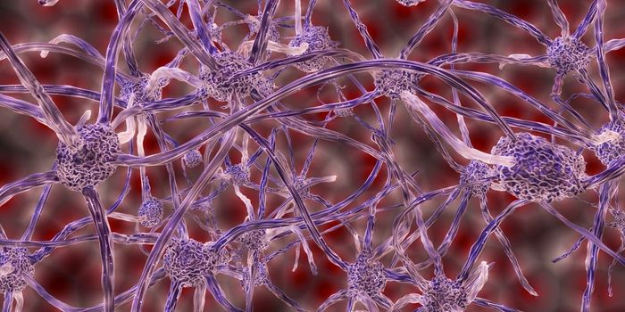
To solve this issue, researchers from Korea developed a noninvasive method to create clear images from scattered infrared light, even after it has passed through a thick layer of bone. Their technique, known as 'laser-scanning reflection-matrix microscopy' (LS-RMM), combines conventional laser-scanning confocal techniques with computational adaptive optics otherwise used to correct optical distortion in ground-based astronomy.
While conventional imaging techniques rely on straight-shooting rays of light (that is often obscured by bone) to build clear, bright images, this new technique is able to interpret both 'straight-shooting' light and those deflected by bone in something known as a 'reflection matrix'. The researchers have already been able to generate high-resolution images of mouse neural networks by using the technique.
A significant step forward in imaging technology, LS-RMM will aid both in the early diagnosis of various diseases and in expediting research in neuroscience. The researchers also note that while LS-RMM requires high amounts of computational power to process aberrations from small, detailed areas, their aberration correction algorithm may also be used by other imaging techniques at less computational cost.
Sources: Science Alert, Nature