The motor cortex homunculus is getting a makeover. New research has essentially redrawn the motor cortex and constitutes one of the most substantial shifts in our topographical knowledge of a sensory brain area in decades. But don’t worry, the big hands and tongue are still on the map.
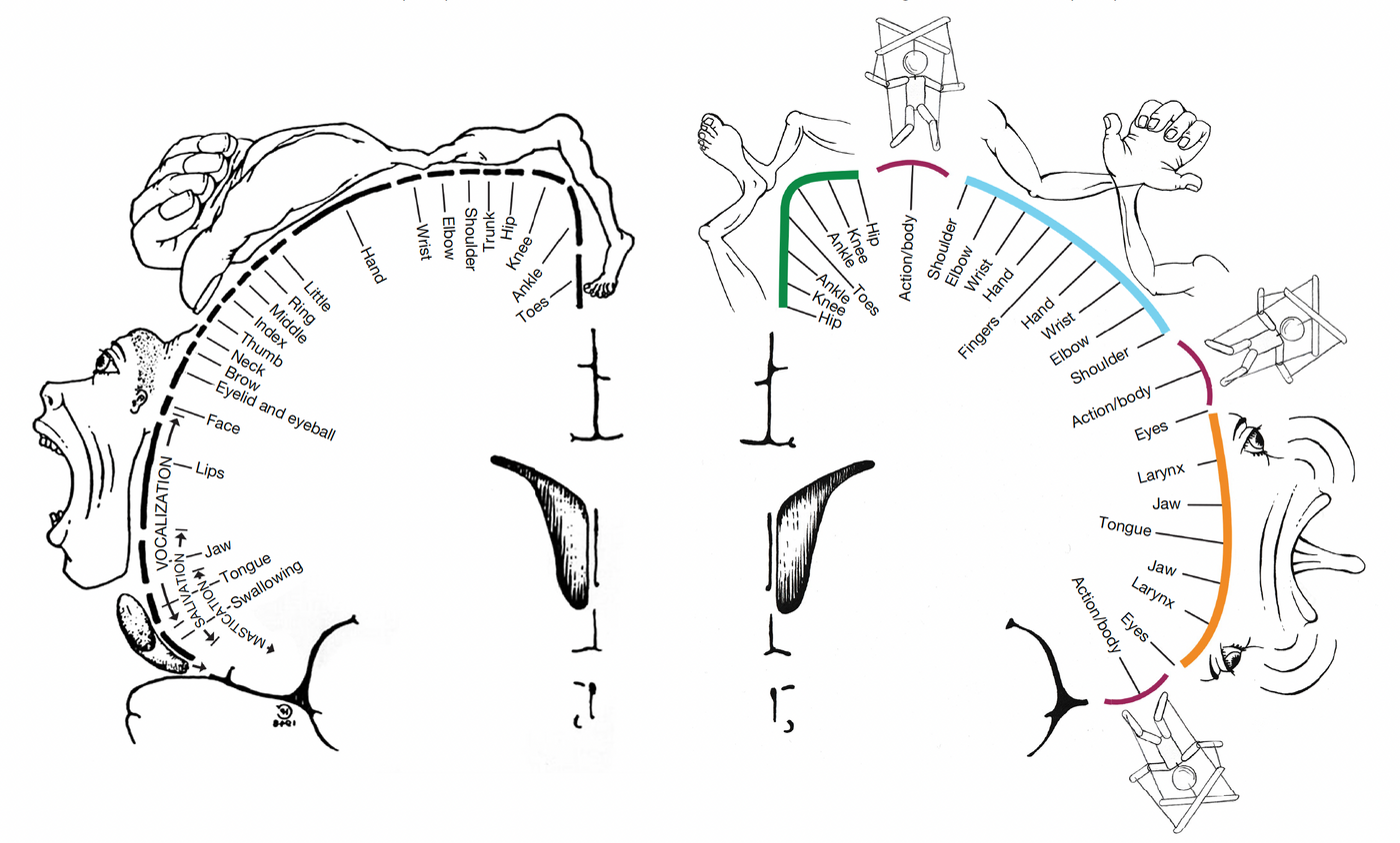
Image Credit: Gordon et al. Nature (2023) adapted from Penfield & Boldrey, E. (1937)
Finding new brain regions seems to be the theme of 2023, with scientific discoveries including new embryotic areas, thalamic slivers, splitting the subarachnoid space, and a thermal cortex.
In the latest shift in our view of the brain’s organization, researchers from the Gordon Neuroimaging Lab at Washington University School of Medicine, St. Louis added previously unidentified areas to the primary motor cortex (M1).
In research published this month in Nature by Gordon et al. and previewed in a bioRxiv publication last year, researchers recorded the activity of the brain using fMRI while participants flexed different muscle groups, performed coordinated movements, produced the noise “eeee” without moving their jaw and compared this to activity during a state of rest (RSFC). Amassing a huge data set allowed researchers to perform detailed brain mapping called precision functional mapping (PFM).
Their first surprise was a connectivity unlike the canonical model of M1. As first author Evan Gordon, Assistant Professor, explains, “the organization of the motor cortex did not correspond well with the “homunculus” described by Wilder Penfield back in the 1930s, which is so well established that it is taught in medical and neuroscience textbooks.”
Gordon et al. shares the story of Penfield, his homunculus, and the misunderstandings that still influence our view of M1. Penfield stimulated points along the vertical length of the M1, mapping which brain areas were responsible for moving what muscle groups. Thus, brain areas were assigned muscle groups like fingers, feet, and even the larynx. But when these areas were artistically drawn along the region of the M1 responsible for them, the result was a disordered row of oddly sized body parts. The homunculus was born.
Even at the time of Penfield, evidence from human electrophysiology experiments suggested there was more to the M1. For example, electrostimulation of the M1 did not always elicit the flexing of a single muscle. Participants reported various unusual feelings, such as the sensation of moving without muscle activity, urges to move against the instructions to hold still, and denying they had just performed an observed movement.
While not wholly incorrect, we now see the limits of Penfield’s model using PFM technology. True to the previous model, large regions of the M1 are dedicated centers for the fine motor movement of the foot, hand, and mouth. These centers still lay along the M1 in the new topography. But Gordon et al. has remapped the role of adjacent areas. As you move along the M1 in either direction away from a center, the surrounding area is dedicated to adjoining body regions. For example, as you move away from the foot center, you enter the ankles region, followed by the knees and hips.
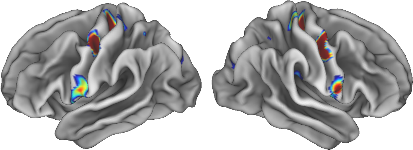
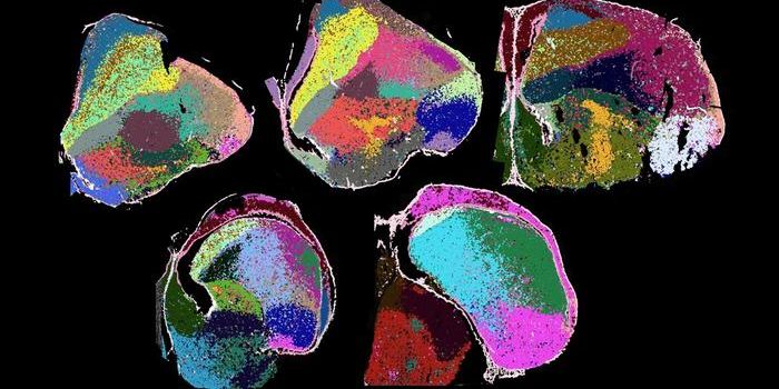
Three colored spots on each half of the brain illuminate special areas in the movement areas of the brain that connect to areas involved in thinking, planning and control of basic bodily functions such as heart rate. The hotter the color, the denser the connections. Researchers at Washington University School of Medicine in St. Louis say that these sites represent a nexus between the body and the mind. Credit: Evan Gordon/Washington University
Even more fascinating is that intermixed between the foot, hand, and mouth regions “lay three areas that were strongly functionally connected to each other... forming a previously unrecognized interdigitated chain down the precentral gyrus,” the Gordon et al. article describes.
These new functional areas, labeled inter-effectors, have unique structures and connections. Responsible for whole-body control, the inter-effectors had lower cortical thickness and higher inter-cortical myelin thickness. But their connection to other regions exposes a previously unappreciated network responsible for coordinating movement.
Gordon describes how the inter-effectors are “connected strongly with a high-level control region of the brain involved in complex planning, setting goals, and monitoring errors,” called the cingulo-opercular network (CON). The CON “is one of the ‘smartest’ areas of the brain, and we’re seeing it connect very strongly with these regions of primary motor cortex.”
This alters our perception of the primary motor cortex's function. “[It] is supposed to only be about moving your body,” says Gordon. “This is the moment when we started thinking of the SCAN.”
The authors dub the network of brain regions associated with the inter-effector regions the somato-cognitive action system (SCAN). This is a “more sophisticated” way for the M1 to behave and “something truly different from the establish body-part-specific areas of motor cortex,” says Gordon.
After it’s make-over, the new M1 contains two behavioral control systems:
- Previously described areas for highly specialized fine movement of our fingers, toes and tongue with less precise adjoining areas
- SCAN: an integrative output system responsible for the coordination of the body as a whole
The new SCAN model is better at explaining the actions of the human electrophysiology experiments from decades earlier. The drive to move or the sensation of moving described by the 20th-century subjects suggests the activity of a system designed to coordinatized goal-directed movement.
The actions of SCAN might also influence other body activities. Findings suggest it integrates information about our autonomic activity or unplanned movements of the digestive organs, lungs, and heart. It also connects to areas of the brain responsible for sensory signals, pain, and decision-making associated with free will (in the dACC).
The large group of researchers completed a range of additional studies, ensuring their findings apply to a variety of future research questions. They aided cross-species studies by reporting detailed scans from macaques. It seems this popular model organism also enjoys effector and inter-effector regions. This is crucial for comparative studies using non-human primates to study complex multi-effector actions.
The actions of SCAN were detectable during infancy and appeared close to adult-like by the age of nine. In one stroke patient, the connections in undamaged areas seemed to be preserved after injury to much of the normal topography.
There’s little doubt this discovery will send medical illustrators back to the drawing board and lead to the recycling of reams of neuroscience textbooks and papers and delete various youtube explanatory videos. Future work will certainly look for similar patterns and details in other sensory regions.
Sources: Nature, bioRxiv, Personal Correspondence with Professor Gordon, Cell (1) (2) (3), Science, Brain, JAMA
Cover photo credit: is licensed under the Creative Commons Attribution-Share Alike 4.0 International license.