Precision is paramount in the field of brain surgery. Neurosurgeons and their teams delicately navigate the brain's intricate circuitry to preserve vital functions while addressing neurological conditions. However, the tools available for mapping functional brain areas have been limited—until now.
A recent breakthrough by researchers at the University of California, San Diego, led by Tchoe et al., introduces an advanced intracranial electroencephalogram (iEEG) microdisplay that promises to revolutionize how we visualize brain activity during surgery.
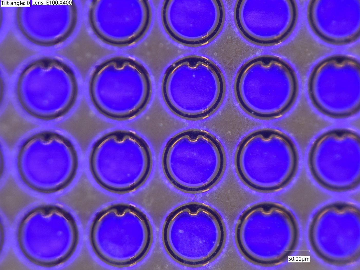
A picture of the microdisplay’s diode grid used with rats. Prof. Shadi Dayeh, UC San Diego (Tchoe et al., 2024)
Navigating the Complexities of Brain Surgery
During brain surgery, precise mapping of functional brain regions is crucial to safeguard critical areas controlling essential functions. Effective communication among neurosurgeons, clinical electrophysiologists, and neurologists is pivotal. Traditionally, this involves verbal interactions to pinpoint crucial brain regions. However, in some surgical settings, vital anatomical information is communicated using antiquated methods, reminiscent of classroom note-passing.
Current electrode grids have limitations, lacking resolution and conforming inadequately to the brain's surface. It’s clear there is a need for innovative solutions to enhance brain mapping accuracy and efficiency during surgery.
Innovative Tool for Real-Time Visualization
Tchoe et al. addressed these challenges by developing an enhanced iEEG microdisplay. This novel technology integrates a micro-electrocorticography electrode grid with an array of light-emitting diodes (LEDs). The result? Real-time visualization of cortical activity directly on the brain's surface during surgery.
Through a series of proof-of-concept experiments in mice and pigs, the researchers demonstrated the microdisplay's capabilities. They successfully visualized cortical activities and identified critical brain landmarks, such as the distinction between the primary sensory and motor cortices. Importantly, the microdisplay's dual-color feature highlighted pathological changes in electrical potentials associated with epileptic activity.
Paving the Way for Future Applications
This pioneering technology heralds a new era in neurosurgical procedures. The iEEG-microdisplay promises to replace traditional methods, such as using paper on the brain's surface for marking resection and preservation areas. Moreover, it opens doors for refining electrophysiological methodologies and advancing neurosurgical techniques.
Looking ahead, the translation of this technology into human applications holds immense promise. The ability to visualize pathological brain activity in real-time during surgery marks a significant leap forward in enhancing surgical precision and patient outcomes.
Sources: Science Translational Medicine