Understanding the Importance of Brain Microstructure Studies
Unlocking the intricate secrets of the human brain's inner workings has long been a pursuit of scientific inquiry. In a groundbreaking study by Shapson-Coe et al., researchers utilized cutting-edge imaging techniques to delve into the brain's microstructure, revealing surprising insights into neuronal connectivity and cellular organization.
The study published in Science delves into a 170-mm-tick slab of human cortex to explore its microarchitecture in high resolution. The aim? To uncover how our brain functions at a cellular and subcellular level.
Utilizing Cutting-Edge Electron Microscopy for High-Resolution Imaging
The researchers employed volume electron microscopy (EM), which allowed for reconstructing every cellular component and synapse. This method capitalizes on the short wavelength of electrons to generate images with nanometer resolution.
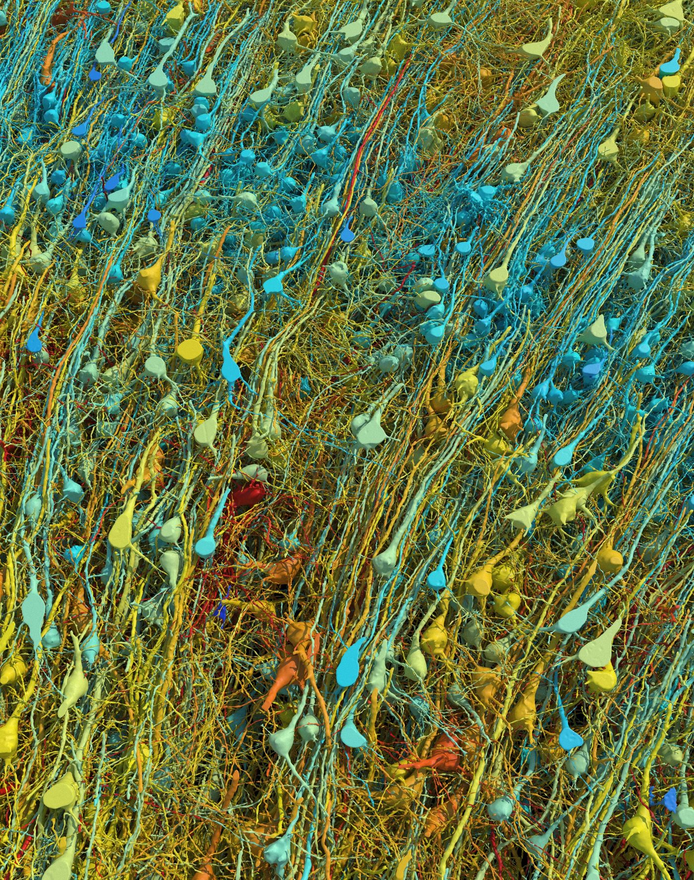
This rendering shows all of the excitatory (pyramidal) neurons in a part of the brain sample, at varying degrees of magnification and tilt. They are colored by size; the cell body (central core) of the cells ranges from 15-30 micrometers across.
Google Research & Lichtman Lab (Harvard University). Renderings by D. Berger (Harvard University)
This study examined a cortical sample from the middle temporal gyrus of a 45-year-old female. This sample, totaling just over one mm^3 in volume, was extracted during surgery to access an underlying epileptic focus in the hippocampus.
Revealing Insights into Neuronal Connectivity and Synapse Classification
The enormity of data gathered from this sample is staggering, amounting to over 1.4 petabytes of digital image data—equivalent to trillions of voxels. This vast dataset enabled visualization of brain tissue at multiple levels—supracellular, cellular, and subcellular—providing insights into the relationships among various cellular elements like neurons, synapses, glia, and blood vessels.
They captured detailed information on their cubic millimeter sample through machine learning, rapid imaging technologies, stand-alone programs, and public proofreading platforms.
The researchers utilized machine learning tools to train automated synapse classifiers. These classifiers identified the components of each synapse and distinguished between excitatory and inhibitory presynaptic terminals.
Through their analysis, several fascinating findings emerged:
- Glial cells outnumber neurons by a ratio of 2:1.
- Oligodendrocytes were identified as the most common cell type.
- Deep-layer excitatory neurons could be classified based on dendritic orientation.
- Rare, powerful axonal inputs were observed composed of up to 50 synaptic connections from a single axon onto a specific neuron
Significance of Rare Axonal Inputs and Synaptic Relationships
Interestingly, while strong connections were rare overall (0.092% of connections), a significant proportion of neurons demonstrated the presence of powerful axonal inputs, suggesting this may be a general characteristic of neuronal innervation in the human cerebral cortex.
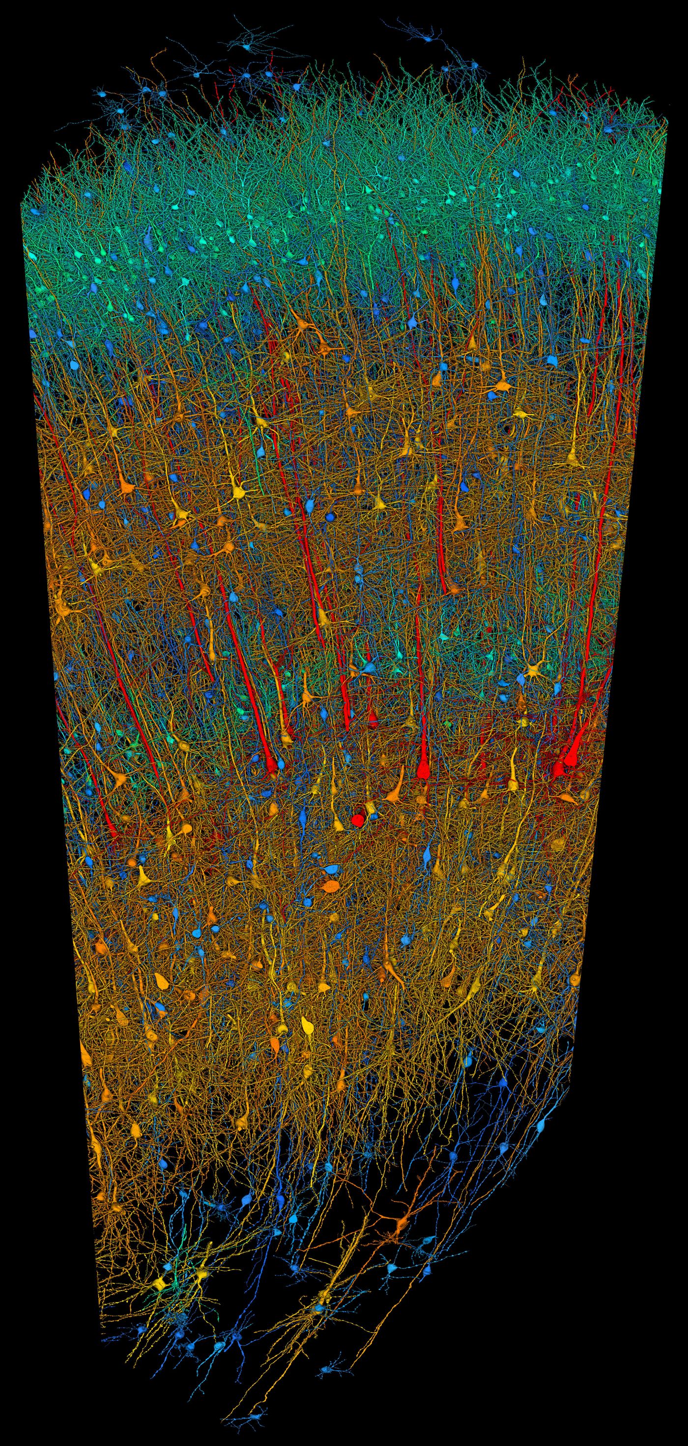
Researchers built a 3D image of nearly every neuron and its connections within a small piece of human brain tissue. Blue neurons are inhibitory neurons. Red, orange, yellow, and green are excitatory neurons colored by size (red is largest, green is smallest) ranging from 15-30 micrometers across at their cores. The sample is approximately 3 mm long. The right image is a closeup of part of the left.
Google Research & Lichtman Lab (Harvard University). Renderings by D. Berger (Harvard University) Google Research & Lichtman Lab (Harvard University). Renderings by D. Berger (Harvard University)
The density of neurons observed in this study (~16,000/mm^3) was notably lower than previously estimated, highlighting differences from other studies and underscoring the need for precise, high-resolution imaging techniques.
Applying Insights from Brain Microstructure Studies
Despite the limitations of the sample being from an individual with epilepsy, this research presents a valuable resource for studying synaptic relationships within the human cortex. The insights gained from this study pave the way for further investigations into the complexities of the human brain, potentially unraveling fundamental mysteries in neuroscience.
In addition to their findings, the researchers have made significant contributions by developing software tools that can aid in further studies, fostering continued exploration into the intricate workings of the human brain.
Sources: Science