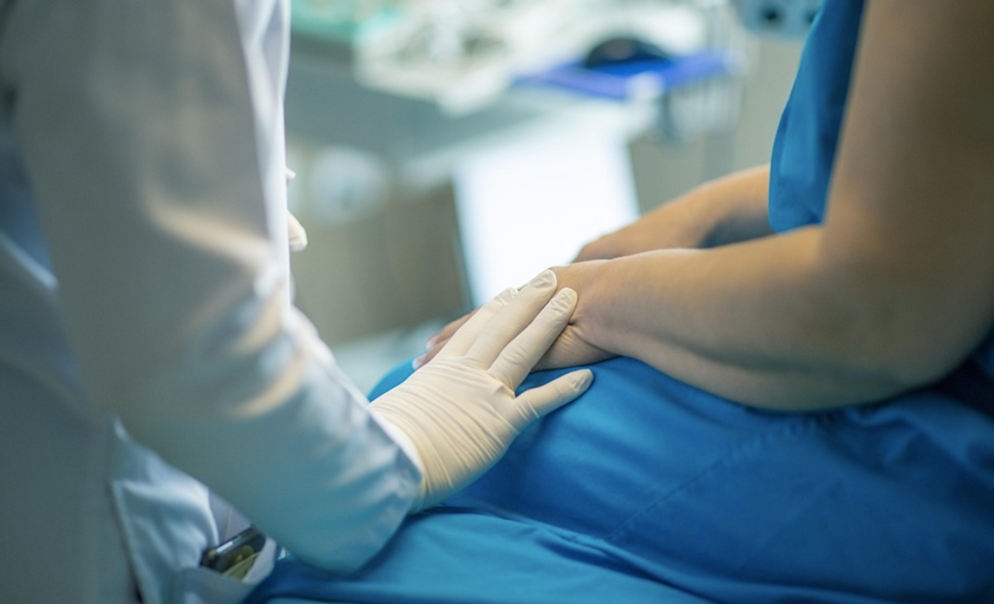
Anyone who needs an unusual mole on their skin checked out may soon get to skip a surgical biopsy, and instead have a virtual biopsy. This tool could be a quick, uninvasive way to identify cancerous cells, as well as reveal any cancerous tissue that might be present and left behind during a surgery. This new tool uses lasers to and generate a three-dimensional reconstruction of cells in a tissue under analysis. Cross-sectional images of that tissue can then be assessed, like slides on a microscope. This work may one day be used not only on skin, but on other parts of the body. The work has been reported in Science Advances.
"We've not only created something that can replace the current gold-standard pathology slides for diagnosing many conditions, but we actually improved the resolution of these scans so much that we start to pick up information that would be extremely hard to see otherwise," said senior study author Adam de la Zerda, PhD, an associate professor of structural biology at Stanford University.
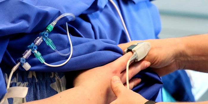
Tissue samples that are collected during a biopsy usually have to be sent to a pathologist, who must cut the tissue into sections that are placed on slides, stain those slides with hematoxylin and eosin (H&E), and evaluate it. This staining process is irreversible, however, and once a tissue is sliced, it cannot be put back together if a different view is needed.
But optical coherence tomography (OCT) scans use lasers to generate a different kind of images of tissue. The technology is currently used to assess the eye. These scans have now been modified and enhanced for use on new organs.
"We kept improving and improving the quality of the image, letting us see smaller and smaller details of a tissue," de la Zerda explained. "And we realized the OCT images we were creating were really getting very similar to the H&Es in terms of what they could show."
However, since clinicians are used to viewing H&E slides, the researchers developed a method to convert the OCT scans into images that resemble them. They relied on computational tools that can now generate multiple images that are similar to H&E from an OCT scan.
"Every physician in a hospital is very much used to reading H&Es, and it was important to us that we translate OCT images into something that physicians were already comfortable with, rather than an entirely new type of image," de la Zerda said.
It may soon be possible to have suspicious moles quickly analyzed with an OCT camera while the patient is in the clinic. The tool may eventually help surgeons ensure that they have removed all of the cancerous tissue from a targeted surgical site; many patients have to return for a second surgery because the first procedure did not remove all of the malignant cells. While more research will be needed before that tool is in use, this work shows that there could be other ways to perform rapid and accurate biopsies to improve patient outcomes.
Experienced research scientist and technical expert with authorships on over 30 peer-reviewed publications, traveler to over 70 countries, published photographer and internationally-exhibited painter, volunteer trained in disaster-response, CPR and DV counseling.